

Protection effect of thymosin β4 on ethanol injury in corneal stromal keratocyte
- PMID: 35062902
- PMCID: PMC8783420
- DOI: 10.1186/s12886-022-02255-8
Protection effect of thymosin β4 on ethanol injury in corneal stromal keratocyte
Abstract
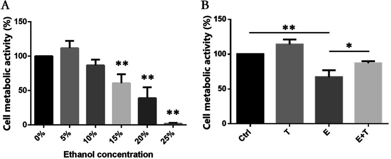
Purpose: To investigate the protective effects of thymosin β4 (Tβ4) on ethanol injured human corneal keratocytes (HCKs).
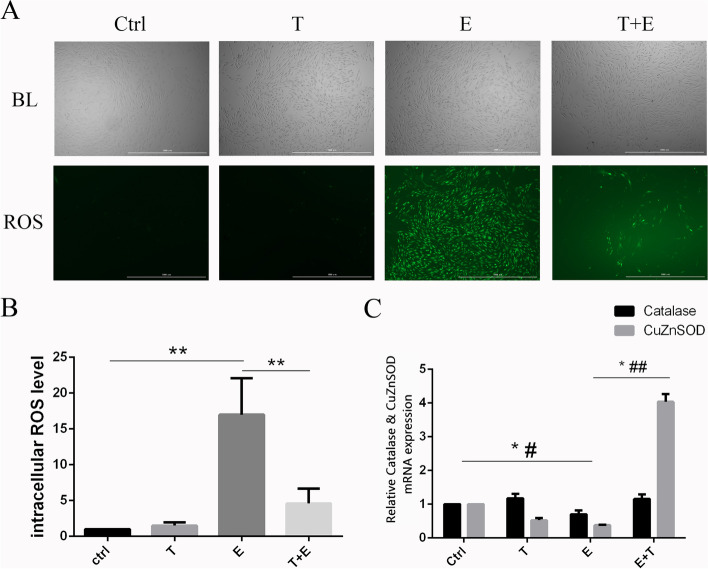
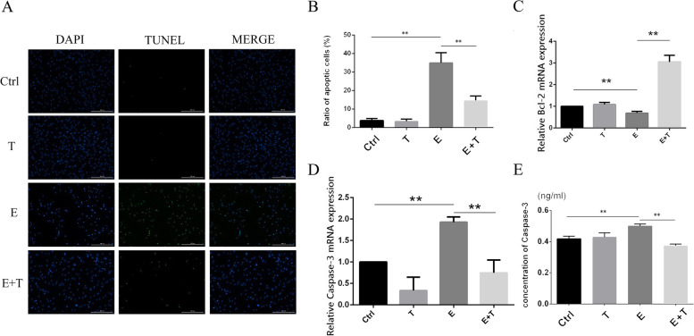
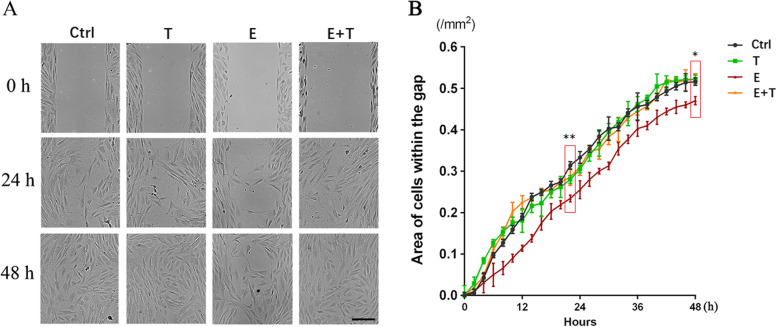
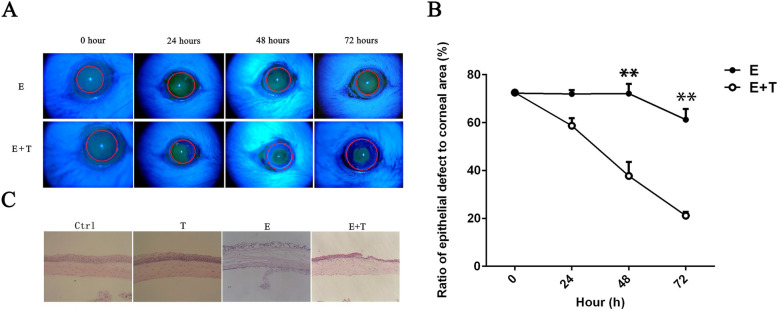
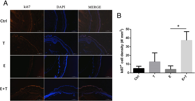
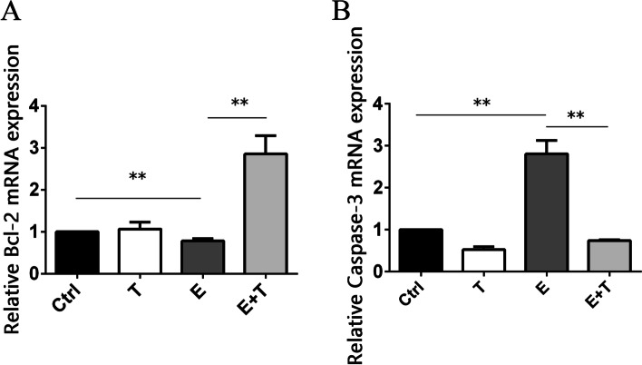
Methods: HCKs and BALB/c mice were chosen as the study subject. Ethanol was used to treat the cells and corneal stroma of mice to build the ethanol injured model in vitro and vivo respectively. CCK-8 was used to evaluate the cell metabolic activity. DCFH-DA was used to detect the intracellular reactive oxygen species level. TUNEL was chose to detect the cell apoptosis rate. The cell proliferation and migration were investigated by using wound healing insert. Wound healing of corneal surface and stroma was observed by using fluorescein sodium eyedrop and HE stain. RT-qPCR, ELISA, and immunostaining were performed to detect gene and protein expression in keratocytes or corneal stroma tissue of mice.
Results: Ethanol induced oxidative stress injury and cell apoptosis on HCKs, and Tβ4 can alleviate it by up-regulating the expression of Bcl-2, catalase, and CuZnSOD, and inhibiting the expression of Caspase-3. Tβ4 promotes the proliferation of HCKs and the process of corneal wound healing. It may relevant to the up-regulated expression of Ki67.
Conclusions: Our study established an ethanol-injured corneal stroma model in both vitro and vivo. The present study confirmed that Tβ4 play a protective effect on the reconstruction process of ethanol-injured corneal stroma.
Keywords: Apoptosis; Cornea stroma; Ethanol; Oxidative stress; Thymosin β4.
© 2022. The Author(s).
Conflict of interest statement
The authors declare that they have no competing interests.
Figures
Similar articles
-
[Protective effect of thymosin β4 against oxidative damage in cultured rabbit corneal epithelial cells].Zhonghua Yan Ke Za Zhi. 2013 Aug;49(8):716-22. Zhonghua Yan Ke Za Zhi. 2013. PMID: 24246811 Chinese.
-
Thymosin-beta4 modulates corneal matrix metalloproteinase levels and polymorphonuclear cell infiltration after alkali injury.Invest Ophthalmol Vis Sci. 2005 Jul;46(7):2388-95. doi: 10.1167/iovs.04-1368. Invest Ophthalmol Vis Sci. 2005. PMID: 15980226
-
Purinergic Signaling Involvement in Thymosin β4-mediated Corneal Epithelial Cell Migration.Curr Eye Res. 2020 Nov;45(11):1352-1358. doi: 10.1080/02713683.2020.1748891. Epub 2020 Apr 2. Curr Eye Res. 2020. PMID: 32223337
-
The corneal fibrosis response to epithelial-stromal injury.Exp Eye Res. 2016 Jan;142:110-8. doi: 10.1016/j.exer.2014.09.012. Exp Eye Res. 2016. PMID: 26675407 Free PMC article. Review.
-
Thymosin beta4 and corneal wound healing: visions of the future.Ann N Y Acad Sci. 2010 Apr;1194:190-8. doi: 10.1111/j.1749-6632.2010.05472.x. Ann N Y Acad Sci. 2010. PMID: 20536468 Review.
Cited by
-
Indomethacin-incorporated microemulsion-laden contact lenses for improved ocular drug delivery and therapeutic efficacy.RSC Adv. 2025 May 14;15(20):16110-16124. doi: 10.1039/d5ra01046b. eCollection 2025 May 12. RSC Adv. 2025. PMID: 40370851 Free PMC article.
References
-
- Seo YS, Kwon JH, Yaqoob U, Yang L, De Assuncao TM, Simonetto DA, Verma VK, Shah VH. HMGB1 recruits hepatic stellate cells and liver endothelial cells to sites of ethanol-induced parenchymal cell injury. Am J Physiol Gastrointest Liver Physiol. 2013;305(11):G838–G848. doi: 10.1152/ajpgi.00151.2013. - DOI - PMC - PubMed
MeSH terms
Substances
LinkOut - more resources
Full Text Sources
Research Materials
