

Extracellular vesicles mediated proinflammatory macrophage phenotype induced by radiotherapy in cervical cancer
- PMID: 35062905
- PMCID: PMC8781113
- DOI: 10.1186/s12885-022-09194-z
Extracellular vesicles mediated proinflammatory macrophage phenotype induced by radiotherapy in cervical cancer
Abstract
Background: Radiotherapy is a highly effective treatment for cervical cancer. Recent studies focused on the radiotherapy induced anti-tumor immunity. Whether tumor-derived extracellular vesicles (EVs) play roles in radiotherapy induced tumor associated macrophage (TAM) polarization remains unclear.
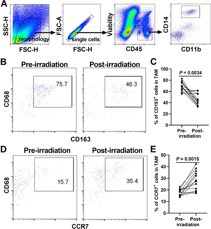
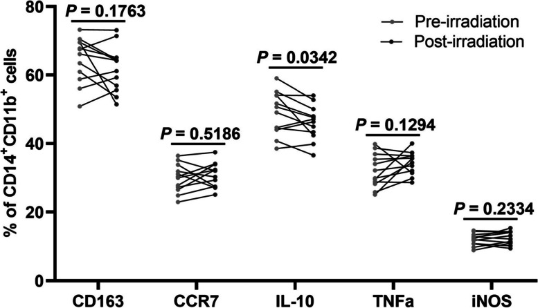
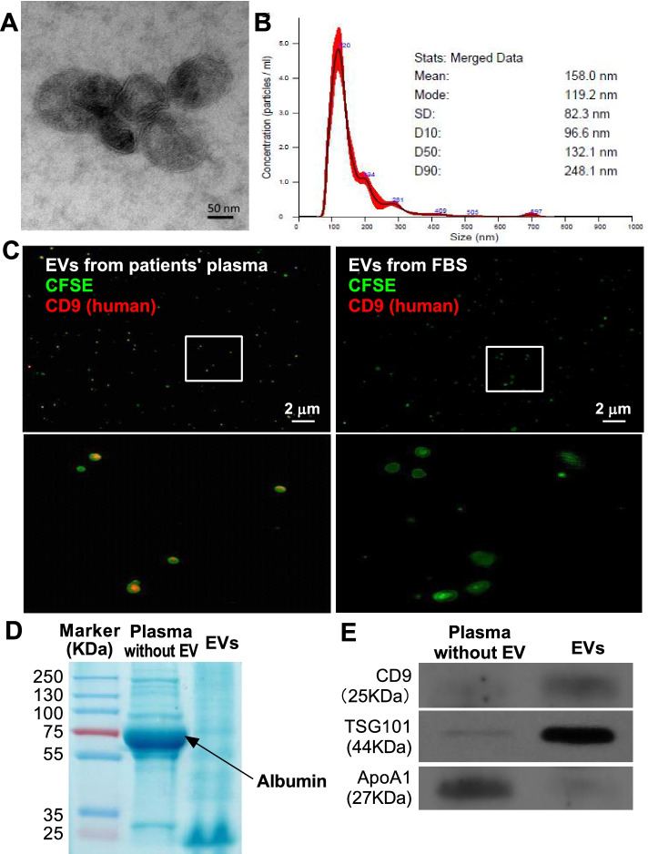
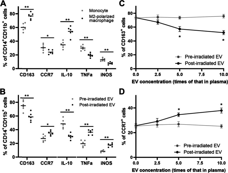
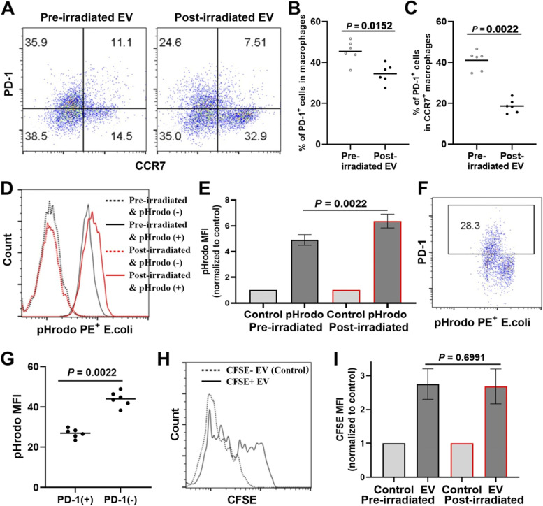
Materials and methods: This study analysed the phenotype of macrophages in cancer tissue and peripheral blood of cervical cancer patients using flow cytometry analysis. The role of EVs from plasma of post-irradiated patients on M2-like transformed macrophages was assessed. The M1- and M2-like macrophages were assessed by expression of cell surface markers (CCR7, CD163) and intracellular cytokines (IL-10, TNFα and iNOS). The capacity of phagocytosis was assessed by PD-1 expression and phagocytosis of pHrodo Red E. coli bioparticles.
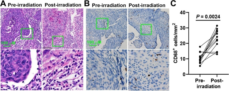
Results: Our results demonstrated that radiotherapy of cervical cancer induced an increase in the number of TAMs and a change in their subtype from the M2-like to the M1-like phenotype (increased expression of CCR7 and decreased expression of CD163). The EVs from plasma of post-irradiated patients facilitated the M2-like to the M1-like phenotype transition (increased expression of CCR7, TNFα and iNOS, and decreased expression of CD163 and IL-10) and increased capacity of phagocytosis (decreased PD-1 expression and increased phagocytosis of pHrodo Red E. coli bioparticles).
Conclusions: Our data demonstrated that irradiation in cervical cancer patients facilitated a proinflammatory macrophage phenotype which could eventually able to mediate anti-tumor immune responses. Our findings highlight the importance of EV in the crosstalk of tumor cells and TAM upon irradiation, which potentially leading to an increased inflammatory response to cancer lesions.
Keywords: Cervical cancer; Extracellular vesicle; Macrophage; Radiotherapy.
© 2022. The Author(s).
Conflict of interest statement
The authors declare that there were no potential conflicts of interest.
Figures
Similar articles
-
Effect of colorectal cancer-derived extracellular vesicles on the immunophenotype and cytokine secretion profile of monocytes and macrophages.Cell Commun Signal. 2018 Apr 24;16(1):17. doi: 10.1186/s12964-018-0229-y. Cell Commun Signal. 2018. PMID: 29690889 Free PMC article.
-
Short-course radiotherapy promotes pro-inflammatory macrophages via extracellular vesicles in human rectal cancer.J Immunother Cancer. 2020 Aug;8(2):e000667. doi: 10.1136/jitc-2020-000667. J Immunother Cancer. 2020. PMID: 32817359 Free PMC article.
-
Tumor cell-released autophagosomes (TRAPs) promote immunosuppression through induction of M2-like macrophages with increased expression of PD-L1.J Immunother Cancer. 2018 Dec 18;6(1):151. doi: 10.1186/s40425-018-0452-5. J Immunother Cancer. 2018. PMID: 30563569 Free PMC article.
-
Crosstalk between extracellular vesicles and tumor-associated macrophage in the tumor microenvironment.Cancer Lett. 2023 Jan 1;552:215979. doi: 10.1016/j.canlet.2022.215979. Epub 2022 Oct 25. Cancer Lett. 2023. PMID: 36306939 Review.
-
Tumor Associated Macrophages in Kidney Cancer.Anal Cell Pathol (Amst). 2016;2016:9307549. doi: 10.1155/2016/9307549. Epub 2016 Oct 11. Anal Cell Pathol (Amst). 2016. PMID: 27807511 Free PMC article. Review.
Cited by
-
Immune modulatory roles of radioimmunotherapy: biological principles and clinical prospects.Front Immunol. 2024 Feb 21;15:1357101. doi: 10.3389/fimmu.2024.1357101. eCollection 2024. Front Immunol. 2024. PMID: 38449871 Free PMC article.
-
Towards Novel Gene and Cell Therapy Approaches for Cervical Cancer.Cancers (Basel). 2022 Dec 30;15(1):263. doi: 10.3390/cancers15010263. Cancers (Basel). 2022. PMID: 36612258 Free PMC article.
-
Identification of Immune and Hypoxia Risk Classifier to Estimate Immune Microenvironment and Prognosis in Cervical Cancer.J Oncol. 2022 Oct 17;2022:6906380. doi: 10.1155/2022/6906380. eCollection 2022. J Oncol. 2022. PMID: 36304989 Free PMC article.
-
Extracellular vesicles and macrophages in tumor microenvironment: Impact on cervical cancer.Heliyon. 2024 Jul 26;10(15):e35063. doi: 10.1016/j.heliyon.2024.e35063. eCollection 2024 Aug 15. Heliyon. 2024. PMID: 39165926 Free PMC article. Review.
-
Influence of Lacidophilin Vaginal Capsules plus rh-IFN-α2b on Efficacy, Vaginal Microecology, and Safety of Patients with HPV Infection.Evid Based Complement Alternat Med. 2022 Aug 3;2022:3632053. doi: 10.1155/2022/3632053. eCollection 2022. Evid Based Complement Alternat Med. 2022. PMID: 35966743 Free PMC article.
References
-
- Bray F, Ferlay J, Soerjomataram I, Siegel RL, Torre LA, Jemal A. Global cancer statistics 2018: GLOBOCAN estimates of incidence and mortality worldwide for 36 cancers in 185 countries. CA Cancer J Clin. 2018;68:394–424. - PubMed
-
- Ronco G, Dillner J, Elfström KM, Tunesi S, Snijders PJ, Arbyn M, et al. Efficacy of HPV-based screening for prevention of invasive cervical cancer: follow-up of four European randomised controlled trials. Lancet. 2014;383:524–532. - PubMed
-
- Smith RA, Andrews KS, Brooks D, Fedewa SA, Manassaram-Baptiste D, Saslow D, Wender RC. Cancer screening in the United States, 2019: A review of current American Cancer Society guidelines and current issues in cancer screening. CA Cancer J Clin. 2019;69:184–210. - PubMed
-
- Joura EA, Giuliano AR, Iversen OE, Bouchard C, Mao C, Mehlsen J, et al. A 9-valent HPV vaccine against infection and intraepithelial neoplasia in women. N Engl J Med. 2015;372:711–723. - PubMed
MeSH terms
Substances
LinkOut - more resources
Full Text Sources
Medical
Research Materials
