Review
. 2022 Jan 21;29(1):6.
doi: 10.1186/s12929-022-00790-6.

LRG1: an emerging player in disease pathogenesis

Affiliations
Review

LRG1: an emerging player in disease pathogenesis

Carlotta Camilli et al. J Biomed Sci. .

Abstract

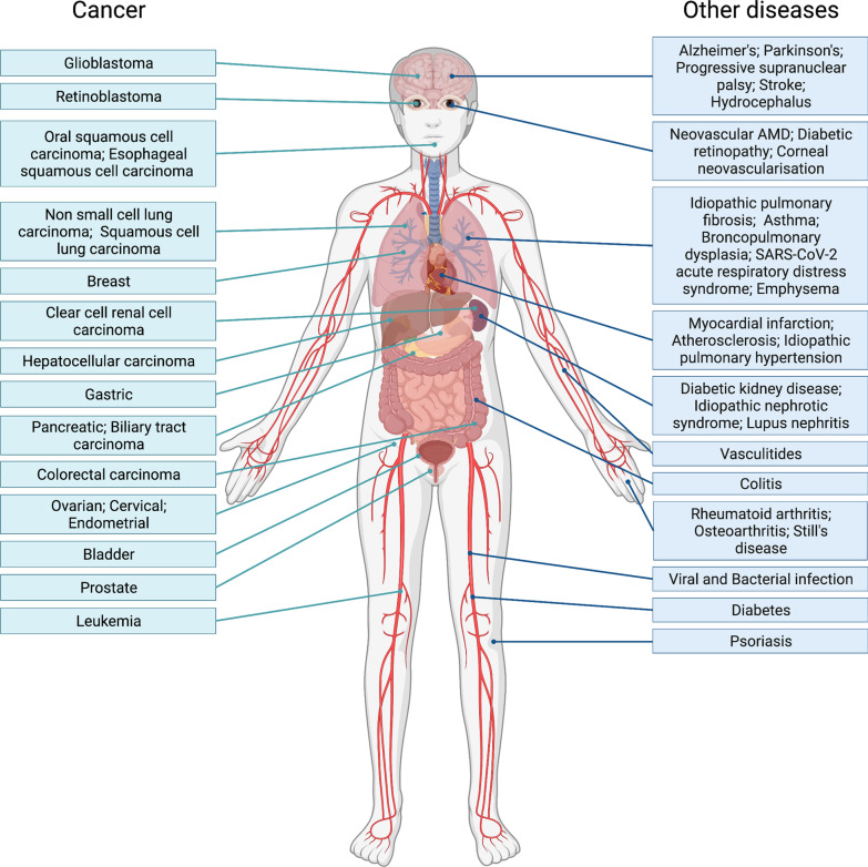
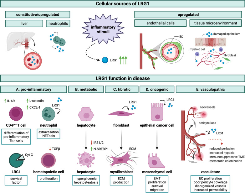
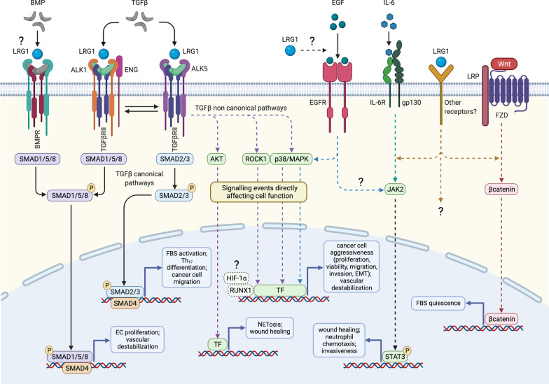
The secreted glycoprotein leucine-rich α-2 glycoprotein 1 (LRG1) was first described as a key player in pathogenic ocular neovascularization almost a decade ago. Since then, an increasing number of publications have reported the involvement of LRG1 in multiple human conditions including cancer, diabetes, cardiovascular disease, neurological disease, and inflammatory disorders. The purpose of this review is to provide, for the first time, a comprehensive overview of the LRG1 literature considering its role in health and disease. Although LRG1 is constitutively expressed by hepatocytes and neutrophils, Lrg1-/- mice show no overt phenotypic abnormality suggesting that LRG1 is essentially redundant in development and homeostasis. However, emerging data are challenging this view by suggesting a novel role for LRG1 in innate immunity and preservation of tissue integrity. While our understanding of beneficial LRG1 functions in physiology remains limited, a consistent body of evidence shows that, in response to various inflammatory stimuli, LRG1 expression is induced and directly contributes to disease pathogenesis. Its potential role as a biomarker for the diagnosis, prognosis and monitoring of multiple conditions is widely discussed while dissecting the mechanisms underlying LRG1 pathogenic functions. Emphasis is given to the role that LRG1 plays as a vasculopathic factor where it disrupts the cellular interactions normally required for the formation and maintenance of mature vessels, thereby indirectly contributing to the establishment of a highly hypoxic and immunosuppressive microenvironment. In addition, LRG1 has also been reported to affect other cell types (including epithelial, immune, mesenchymal and cancer cells) mostly by modulating the TGFβ signalling pathway in a context-dependent manner. Crucially, animal studies have shown that LRG1 inhibition, through gene deletion or a function-blocking antibody, is sufficient to attenuate disease progression. In view of this, and taking into consideration its role as an upstream modifier of TGFβ signalling, LRG1 is suggested as a potentially important therapeutic target. While further investigations are needed to fill gaps in our current understanding of LRG1 function, the studies reviewed here confirm LRG1 as a pleiotropic and pathogenic signalling molecule providing a strong rationale for its use in the clinic as a biomarker and therapeutic target.

Keywords: Cancer; Diabetes; Endothelial cell; Fibrosis; Immunity; Inflammation; LRG1; Neovascularization; Neutrophils; Vascular normalization.


Conflict of interest statement

JG and SEM are founders and members of the scientific advisory board of a company spun out by UCL Business to commercialize a LRG1 function-blocking therapeutic antibody developed through the UK Medical Research Council DPFS funding scheme. JG and SEM are shareholders of this company and named inventors on three patents related to LRG1 as a therapeutic target.

Figures

Fig. 1
Fig. 1
LRG1 expression in disease. Human diseases characterized by the upregulation of LRG1 expression. These illustrate the pleiotropic and wide-spanning role LRG1 plays in disease. Considerable data now show that LRG1 is a contributing factor to the disease process and not simply a response to the condition
Fig. 2
Fig. 2
LRG1 protein structure. Schematic representation of LRG1 protein structure. A LRG1 is a 312 aa protein which contains 8 leucine-rich repeats (LRR), 4 N-linked, 1 O-linked glycosylation sites and 2 disulphide bonds. Upon cleavage of the N-terminal signal peptide, LRG1 is released in the extracellular space. The mature form, around 50 kDa, may vary in weight depending on the glycosylation pattern and multimer formation. B LRG1 structure as predicted by ALPHAFOLD2 through deep learning algorithms [223]. β-sheet in green, α-helix in red
Fig. 3
Fig. 3
LRG1 expression in normal and cancer tissues. A A polyclonal (Proteintech) or monoclonal (Magacizumab) antibody was used for the detection of human LRG1 (brown) respectively in human liver (left, scale bar: 250 µm) and testis (right, scale bar: 100 µm). B Immunohistochemistry showing upregulation of LRG1 expression (brown) in human breast cancer (right) compared to healthy control (scale bar: 60–62 µm). The arrow indicates an example of LRG1pos blood vessel. C Lrg1 mRNA (green) detected by RNA scope and immunofluorescence for Collagen IV (white) profiling tissue vessels in normal mouse liver (scale bar: 100 µm). D Lrg1 mRNA (red) detected by RNA scope and immunofluorescence for Collagen IV (white) profiling tissue vessels in normal mouse lung (scale bar: 100 µm). The arrow points to Lrg1 mRNA expressed by lung alveolar epithelium while the box shows details of additional LRG1pos stromal cells. E An anti-human polyclonal antibody (Proteintech) was used for the detection of LRG1 (red) in normal human lung (scale bar: 100 µm). F Upregulation of LRG1 expression in murine metastatic lung tumours. Top: examples of Lrg1 mRNA detected by RNA scope (green) and Collagen IV stained by immunofluorescence (white); left: low magnification of metastatic tumour mass (scale bar: 100 µm); middle: Lrg1 expression by cancer cells or cancer-associated fibroblasts (scale bar: 50 µm); right: Lrg1 expression by tumour vessels (scale bar: 50 µm). Bottom left: Lrg1 mRNA (green) detected by RNA scope and immunofluorescence for Podoplanin (red) (scale bar: 100 µm); right: Lrg1 mRNA detected by RNA scope (green) and immunofluorescence for the endothelial markers ERG (red) and Podocalyxin (white) (scale bar: 50 µm). Murine metastatic tumour samples were kindly provided by M. Singhal and H. G. Augustine, Heidelberg University, Germany. Human samples were purchased from Biomax and Covance.
Fig. 4
Fig. 4
Regulation of Lrg1 expression. Schematic representation of the mechanisms regulating LRG1 expression at transcriptional and post-transcriptional levels. Several pro-inflammatory signalling molecules, including cytokines and bacteria-derived LPS, drive the expression of Lrg1 by promoting the activation of different transcription factors in a cell- and context-specific manner. Importantly, the combined stimulation with different cytokines has a synergistic effect on the activity of Lrg1 promoter. Biomechanical forces also stimulate Lrg1 expression through the FAK/ERK/ELK1 axis. Various non-coding RNAs have been associated with Lrg1 regulation. While the lncRNA TUG1 directly facilitates Lrg1 transcription, miR-335, miR-494, miR-497, miR-150-5p and miR-24-3p promote the degradation of Lrg1 mRNA and therefore are often downregulated in cancer. TGFβ-induced methylation has also been reported to favour expression of the Lrg1 gene. Finally, LRG1 protein is differentially glycosylated in a cell- and function-specific fashion prior to secretion into the extracellular space. OSM Osteopontin, lncRNA long non-coding RNA
Fig. 5
Fig. 5
LRG1 functions in disease progression. Schematic representation of LRG1 cell sources and pathological functions. Following various inflammatory stimuli, including infection, injury, autoimmune disease, and tumour-associated inflammation, LRG1 may be produced systemically and/or at the local tissue level. Predominant cellular sources include hepatocytes, neutrophils, and endothelial cells but also other components of the tissue microenvironment, namely epithelial cells, fibroblasts, and other types of myeloid cells. LRG1 pathogenic functions may be initiated through autocrine and paracrine activity and can be broadly classified into A pro-inflammatory: LRG1 favours immune cell participation at the inflammatory site by (i) counteracting TGFβ-driven anti-proliferative function on hematopoietic progenitors; (ii) promoting the extravasation and activation of neutrophils; and (iii) enhancing the differentiation of naïve CD4pos T cells into pro-inflammatory Th17 lymphocytes. Additionally, LRG1 acts as a survival factor for circulating immune cells by neutralizing Cyt c cytotoxicity. B metabolic: LRG1 affects hepatocytes by suppressing fatty acid catabolism, promoting lipogenesis through activation of SREBP1, and inhibiting the expression of IRS1/2 thus contributing to hepatosteatosis and hyperglycemia. C fibrotic: LRG1 promotes the functional transition of fibroblasts (and epithelial cells, not shown) into ECM-producing cells in fibrosis. D oncogenic: LRG1 contributes to cancer cell malignancy by promoting EMT and exerting proliferative and anti-apoptotic functions. E vasculopathic: LRG1 affects vessel stability by promoting dysfunctional angiogenesis and interfering with EC-pericyte crosstalk. These effects contribute to the formation of disorganized and highly permeable capillaries. Notably, these outcomes indirectly sustain and amplify some of the direct effects, as dysfunctional and poorly perfused vessels are responsible for the establishment of a highly hypoxic microenvironment which, in turn, contributes to fibrosis, immunosuppression and cancer cell aggressiveness. Cyt c cytochrome c, IRS insulin receptor substrate, N-SREBP1 nuclear sterol regulatory element binding protein 1, ECM extracellular matrix, EMT epithelial-mesenchymal transition, EC endothelial cell
Fig. 6
Fig. 6
LRG1 signalling pathways. Simplified schematic representation of generic signalling pathways known, or speculated, to be modulated by LRG1 in a cell-specific manner. LRG1 likely modifies cell behaviour both directly, by altering the cell transcriptome, and indirectly by interfering with intermediate steps of the signalling cascades. LRG1 has been mainly described as a modifier of the TGFβ canonical pathway. While promoting pathogenic angiogenesis in endothelial cells through the ALK1-Smad1/5/8 pathway [4, 23], LRG1 may also modulate the ALK5-Smad2/3 arm to favour the formation of myofibroblasts [16, 68] and Th17 lymphocytes [27], thus sustaining fibrosis and inflammation, as well as glioma cell migration [194]. TGFβ non-canonical signalling is also likely to mediate some of the LRG1-driven biological functions including neutrophil activation and wound healing via AKT [56, 64], as well as the modulation of stem/cancer cell phenotype via ROCK1 [39] and p38/MAPK [31, 87]. Activation of the TGFβ-related transcription factors HIF-1α [197] and RUNX1 [50] has also been associated with LRG1 pro-oncogenic functions, although the specific upstream pathways subject to LRG1 modification in this context remain to be formally clarified. Additional transduction factors involved in LRG1 signalling include (i) EGFR which promotes pancreatic cancer cell malignancy through p38/MAPK [49], dissemination of melanoma cells [90] and cornea repair through STAT3 [64]; (ii) the IL-6/STAT3 axis which modulates neutrophil chemotaxis [57]; (iii) Wnt/βcatenin which, in the heart, inhibit fibroblast proliferation and migration [37]. Further investigations are needed to address whether LRG1 modulates the activity of other receptor-mediated signalling pathways including BMPs, and whether other receptors may also be directly or indirectly affected by LRG1. BMP bone morphogenic protein, EC endothelial cell, FBS fibroblast, EMT epithelial-mesenchymal transition, FZD Frizzled, TF transcription factor, NET neutrophil extracellular trap

References

    1. Haupt H, Baudner S. Isolation and characterization of an unknown, leucine-rich 3.1-S-alpha2-glycoprotein from human serum (author's transl) Hoppe Seylers Z Physiol Chem. 1977;358(6):639–646. - PubMed
    1. Kobe B, Kajava AV. The leucine-rich repeat as a protein recognition motif. Curr Opin Struct Biol. 2001;11(6):725–732. - PubMed
    1. Ng A, Xavier RJ. Leucine-rich repeat (LRR) proteins: integrators of pattern recognition and signaling in immunity. Autophagy. 2011;7(9):1082–1084. - PMC - PubMed
    1. Wang X, Abraham S, McKenzie JAG, Jeffs N, Swire M, Tripathi VB, et al. LRG1 promotes angiogenesis by modulating endothelial TGF-β signalling. Nature. 2013;499(7458):306–311. - PMC - PubMed
    1. Kallenberg D, Tripathi V, Javaid F, Pilotti C, George J, Davis S, et al. A Humanized Antibody against LRG1 that Inhibits Angiogenesis and Reduces Retinal Vascular Leakage. BioRxiv. 2020;55:137.