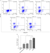
Antigenic and immunogenic properties of recombinant proteins consisting of two immunodominant African swine fever virus proteins fused with bacterial lipoprotein OprI
- PMID: 35062983
- PMCID: PMC8781047
- DOI: 10.1186/s12985-022-01747-9
Antigenic and immunogenic properties of recombinant proteins consisting of two immunodominant African swine fever virus proteins fused with bacterial lipoprotein OprI
Abstract
Background: African swine fever (ASF) is a highly fatal swine disease, which threatens the global pig industry. There is no commercially available vaccine against ASF and effective subunit vaccines would represent a real breakthrough.
Methods: In this study, we expressed and purified two recombinant fusion proteins, OPM (OprI-p30-modified p54) and OPMT (OprI-p30-modified p54-T cell epitope), which combine the bacterial lipoprotein OprI with ASF virus proteins p30 and p54. Purified recombinant p30 and modified p54 expressed alone or fused served as controls. The activation of dendritic cells (DCs) by these proteins was first assessed. Then, humoral and cellular immunity induced by the proteins were evaluated in mice.
Results: Both OPM and OPMT activated DCs with elevated expression of relevant surface molecules and proinflammatory cytokines. Furthermore, OPMT elicited the highest levels of antigen-specific IgG responses, cytokines including interleukin-2, interferon-γ, and tumor necrosis factor-α, and proliferation of lymphocytes. Importantly, the sera from mice vaccinated with OPM or OPMT neutralized more than 86% of ASF virus in vitro.
Conclusions: Our results suggest that OPMT has good immunostimulatory activities and immunogenicity in mice, and might be an appropriate candidate to elicit immune responses in swine. Our study provides valuable information on further development of a subunit vaccine against ASF.
Keywords: African swine fever virus; Immune response; Immunomodulation; OprI; Recombinant fusion proteins; Vaccine.
© 2022. The Author(s).
Conflict of interest statement
The authors declare no conflict of interests.
Figures


Similar articles
-
Evaluation of humoral and cellular immune responses induced by a cocktail of recombinant African swine fever virus antigens fused with OprI in domestic pigs.Virol J. 2023 May 26;20(1):104. doi: 10.1186/s12985-023-02070-7. Virol J. 2023. PMID: 37237390 Free PMC article.
-
Antigenicity and immunogenicity of recombinant proteins comprising African swine fever virus proteins p30 and p54 fused to a cell-penetrating peptide.Int Immunopharmacol. 2021 Dec;101(Pt A):108251. doi: 10.1016/j.intimp.2021.108251. Epub 2021 Oct 29. Int Immunopharmacol. 2021. PMID: 34715492
-
The African swine fever virus proteins p54 and p30 are involved in two distinct steps of virus attachment and both contribute to the antibody-mediated protective immune response.Virology. 1998 Apr 10;243(2):461-71. doi: 10.1006/viro.1998.9068. Virology. 1998. PMID: 9568043
-
African Swine Fever Virus Biology and Vaccine Approaches.Adv Virus Res. 2018;100:41-74. doi: 10.1016/bs.aivir.2017.10.002. Epub 2017 Nov 21. Adv Virus Res. 2018. PMID: 29551143 Review.
-
Multifaceted Immune Responses to African Swine Fever Virus: Implications for Vaccine Development.Vet Microbiol. 2020 Oct;249:108832. doi: 10.1016/j.vetmic.2020.108832. Epub 2020 Aug 29. Vet Microbiol. 2020. PMID: 32932135 Review.
Cited by
-
Evaluation of humoral and cellular immune responses induced by a cocktail of recombinant African swine fever virus antigens fused with OprI in domestic pigs.Virol J. 2023 May 26;20(1):104. doi: 10.1186/s12985-023-02070-7. Virol J. 2023. PMID: 37237390 Free PMC article.
-
Vaccines for African swine fever: an update.Front Microbiol. 2023 Apr 27;14:1139494. doi: 10.3389/fmicb.2023.1139494. eCollection 2023. Front Microbiol. 2023. PMID: 37180260 Free PMC article. Review.
-
Designing a multi-epitope vaccine against African swine fever virus using immunoinformatics approach.Sci Rep. 2025 May 8;15(1):16044. doi: 10.1038/s41598-025-00705-z. Sci Rep. 2025. PMID: 40341420 Free PMC article.
-
Summary of the Current Status of African Swine Fever Vaccine Development in China.Vaccines (Basel). 2023 Mar 29;11(4):762. doi: 10.3390/vaccines11040762. Vaccines (Basel). 2023. PMID: 37112673 Free PMC article.
-
Advancement in the development of gene/protein-based vaccines against African swine fever virus.Curr Res Microb Sci. 2024 Mar 12;6:100232. doi: 10.1016/j.crmicr.2024.100232. eCollection 2024. Curr Res Microb Sci. 2024. PMID: 38545259 Free PMC article. Review.
References
-
- Blome S, Franzke K, Beer M. African swine fever - A review of current knowledge. Virus Res. 2020;287:198099. - PubMed
-
- Revilla Y, Pérez-Núñez D, Richt JA. African swine fever virus biology and vaccine approaches. Adv Virus Res. 2018;100:41–74. - PubMed
-
- Alonso C, Borca M, Dixon L, Revilla Y, Rodriguez F, Escribano JM, Ictv Report C. ICTV virus taxonomy profile: asfarviridae. J Gen Virol. 2018;99:613–614. - PubMed
Publication types
MeSH terms
Substances
LinkOut - more resources
Full Text Sources

