

Review
doi: 10.1016/j.molcel.2021.12.024.
The new era of quantitative cell imaging-challenges and opportunities
Affiliations
- PMID: 35063094
- PMCID: PMC10339817
- DOI: 10.1016/j.molcel.2021.12.024
Item in Clipboard
Review
The new era of quantitative cell imaging-challenges and opportunities
Mol Cell.
.
Abstract
Quantitative optical microscopy-an emerging, transformative approach to single-cell biology-has seen dramatic methodological advancements over the past few years. However, its impact has been hampered by challenges in the areas of data generation, management, and analysis. Here we outline these technical and cultural challenges and provide our perspective on the trajectory of this field, ushering in a new era of quantitative, data-driven microscopy. We also contrast it to the three decades of enormous advances in the field of genomics that have significantly enhanced the reproducibility and wider adoption of a plethora of genomic approaches.
Copyright © 2021 Elsevier Inc. All rights reserved.
Figures
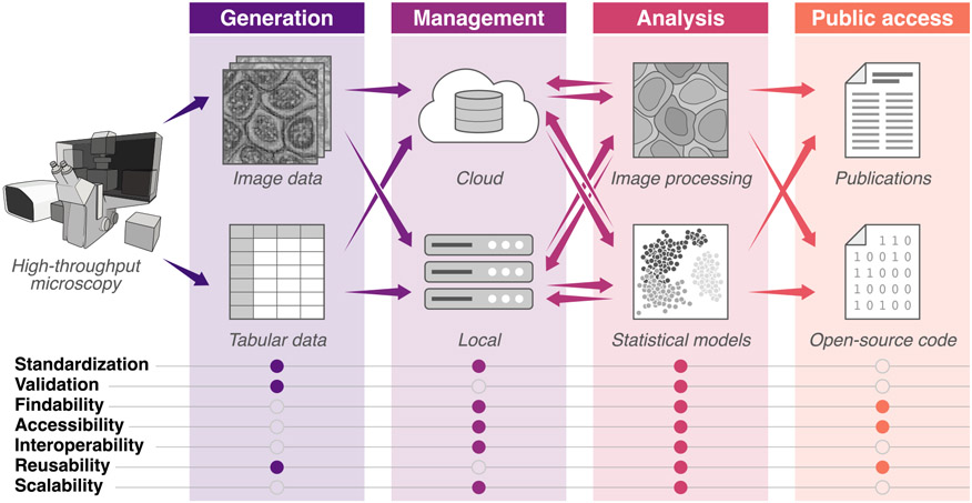
This illustration highlights ways in which the community can build consensus, incentives, and use cases across the imaging pipeline. Information transfer, from data generation through publication, is shown with arrows. Addressing the list of features in filled circles will enable the iterative sharing of data, resources, and tools in robust and easy-to-access repositories, which is critical to the growth of the quantitative cell imaging community and closes the loop between analysis, generation, and discovery.
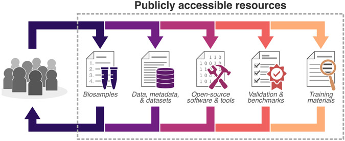
The consistent use of dynamic standards, benchmarks, and annotations for data generation and the sharing of these resources in repositories is critical to developing a quantitative cell imaging community. Supporting publicly available resources positions the community to build upon shared expertise and develop resources that will empower and engage a new wave of investigators and lower the barrier for productive collaborations.
References
-
- Anderson N, and Badano A (2016). Technical Performance Assessment of Digital Pathology Whole Slide Imaging Devices - Guidance for Industry and Food and Drug Administration Staff (U.S. Department of Health and Human Services, Food and Drug Administration; ).
-
- Bajcsy P, Chalfoun J, and Simon M (2018). Web Microanalysis of Big Image Data (Springer; ).
-
- Betzig E, Patterson GH, Sougrat R, Lindwasser OW, Olenych S, Bonifacino JS, Davidson MW, Lippincott-Schwartz J, and Hess HF (2006). Imaging intracellular fluorescent proteins at nanometer resolution. Science 313, 1642–1645. - PubMed
-
- Bickle M. (2010). The beautiful cell: high-content screening in drug discovery. Anal. Bioanal. Chem 393, 219–226. - PubMed
Publication types
MeSH terms
Grants and funding
LinkOut - more resources
Full Text Sources
Miscellaneous
