. 2022 Mar;298(3):101608.
doi: 10.1016/j.jbc.2022.101608. Epub 2022 Jan 20.

Targeting Wnt/tenascin C-mediated cross talk between pancreatic cancer cells and stellate cells via activation of the metastasis suppressor NDRG1

Affiliations

Targeting Wnt/tenascin C-mediated cross talk between pancreatic cancer cells and stellate cells via activation of the metastasis suppressor NDRG1

Bekesho Geleta et al. J Biol Chem. 2022 Mar.

Abstract

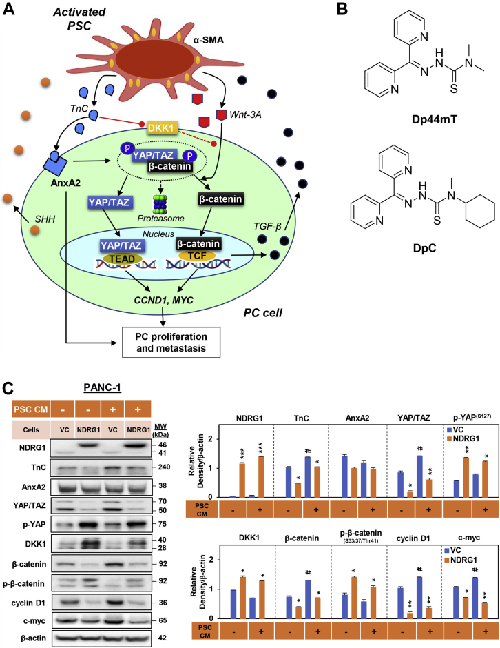
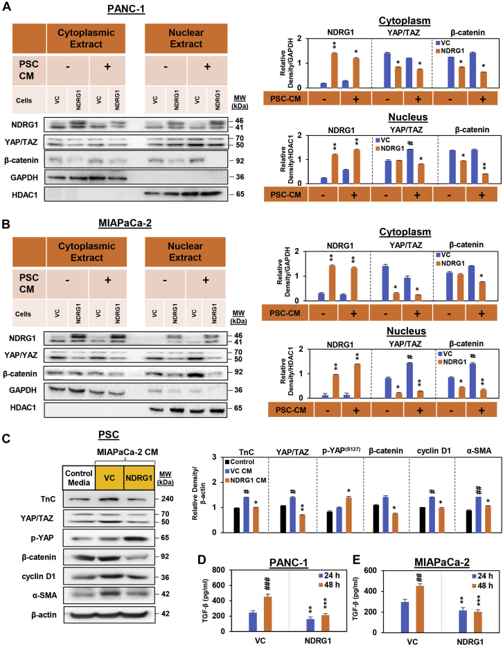
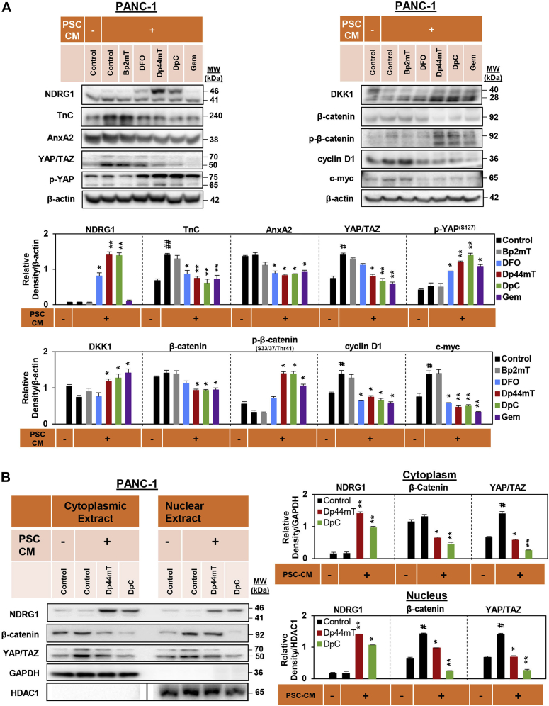
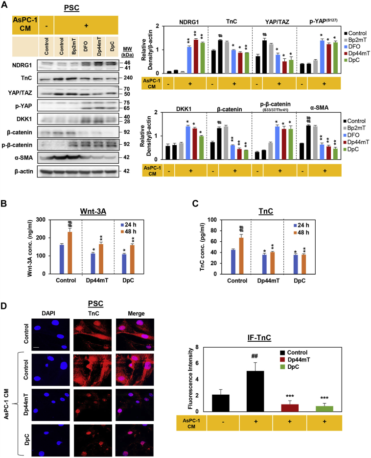
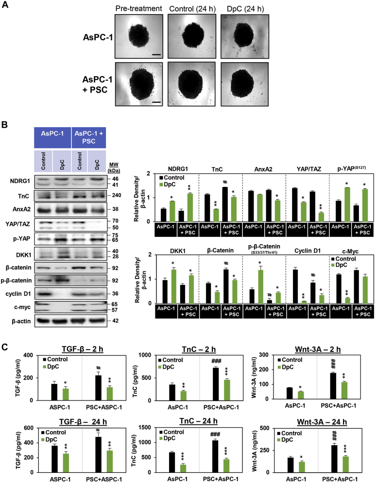
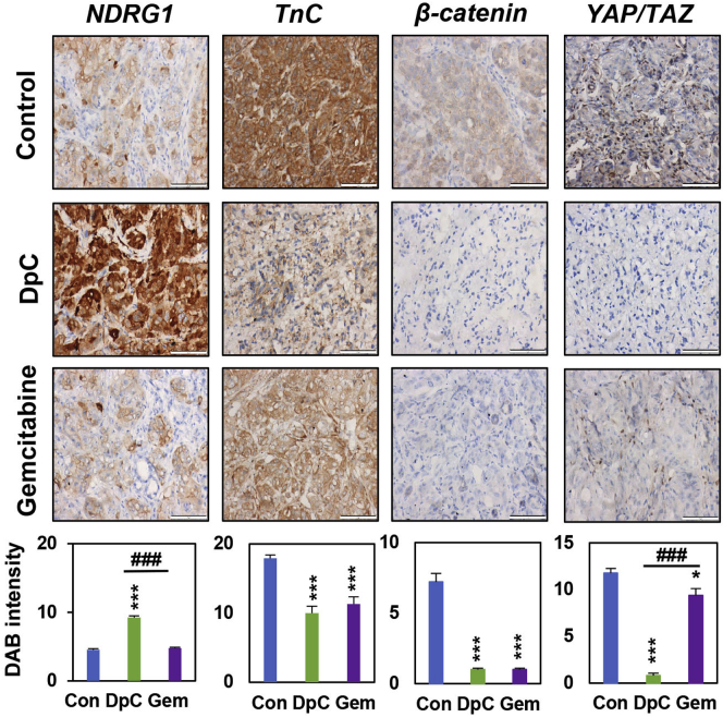
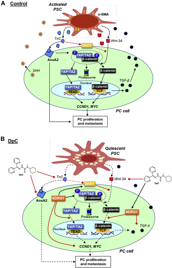
A major barrier to successful pancreatic cancer (PC) treatment is the surrounding stroma, which secretes growth factors/cytokines that promote PC progression. Wnt and tenascin C (TnC) are key ligands secreted by stromal pancreatic stellate cells (PSCs) that then act on PC cells in a paracrine manner to activate the oncogenic β-catenin and YAP/TAZ signaling pathways. Therefore, therapies targeting oncogenic Wnt/TnC cross talk between PC cells and PSCs constitute a promising new therapeutic approach for PC treatment. The metastasis suppressor N-myc downstream-regulated gene-1 (NDRG1) inhibits tumor progression and metastasis in numerous cancers, including PC. We demonstrate herein that targeting NDRG1 using the clinically trialed anticancer agent di-2-pyridylketone-4-cyclohexyl-4-methyl-3-thiosemicarbazone (DpC) inhibited Wnt/TnC-mediated interactions between PC cells and the surrounding PSCs. Mechanistically, NDRG1 and DpC markedly inhibit secretion of Wnt3a and TnC by PSCs, while also attenuating Wnt/β-catenin and YAP/TAZ activation and downstream signaling in PC cells. This antioncogenic activity was mediated by direct inhibition of β-catenin and YAP/TAZ nuclear localization and by increasing the Wnt inhibitor, DKK1. Expression of NDRG1 also inhibited transforming growth factor (TGF)-β secretion by PC cells, a key mechanism by which PC cells activate PSCs. Using an in vivo orthotopic PC mouse model, we show DpC downregulated β-catenin, TnC, and YAP/TAZ, while potently increasing NDRG1 expression in PC tumors. We conclude that NDRG1 and DpC inhibit Wnt/TnC-mediated interactions between PC cells and PSCs. These results further illuminate the antioncogenic mechanism of NDRG1 and the potential of targeting this metastasis suppressor to overcome the oncogenic effects of the PC-PSC interaction.

Keywords: NDRG1; TnC; Wnt; pancreatic cancer; tumor microenvironment.


Conflict of interest statement

Conflict of interest The authors declare that they have no conflicts of interest with the contents of this article.

Figures

Figure 1
Figure 1
NDRG1 expression reduced TnC, YAP/TAZ, β-catenin, cyclin D1,and c-myc in PANC-1 cells.A, schematic of the oncogenic signaling pathways involved in pancreatic cancer cell (PC) and pancreatic stellate cell (PSC) bidirectional oncogenic signaling. Activated PSCs induce β-catenin and YAP/TAZ signaling in PC cells. Activation of PSCs results in increased TnC secretion, which acts on PC cells to activate AnxA2 and promote PC proliferation and metastasis. In addition, TnC downregulates DKK1 promoter activity to decrease the expression of this Wnt antagonist. This leads to dissociation of the cytoplasmic β-catenin destruction complex, enabling activation of both β-catenin and YAP/TAZ signaling. Once released from the destruction complex, β-catenin and YAP/TAZ translocate to the nucleus inducing CCND1 and MYC expression, which promotes PC progression. PC cells activate PSCs by secreting sonic hedgehog (SHH) (31, 32, 33) and transforming growth factor-β (TGF-β) that promotes oncogenic activation of PSCs, enabling increased production of TnC and ligands including Wnt-3A. These interactions are part of the oncogenic, bidirectional cross talk between PC and PSC cells. B, line drawings of the structures of Dp44mT and DpC. C, Western blot analysis of PANC-1 pancreatic cancer cells examining the effect of NDRG1 overexpression versus the vector control (VC) on the levels of NDRG1, TnC, AnxA2, YAP/TAZ, p-YAP, DKK1, β-catenin, p-β-catenin, cyclin D1, and c-myc after a 24 h incubation in the presence or absence of PSC-conditioned medium (PSC CM). β-actin was used as a protein-loading control. Blots are representative of three independent experiments. Densitometry results are mean ± SD (n = 3 experiments). ∗p < 0.05; ∗∗p < 0.01; ∗∗∗p < 0.001 denote statistical significance comparing NDRG1 overexpression to the respective VC. #p < 0.05 comparing PSC CM treatment with the respective control medium-treated cells.
Figure 2
Figure 2
NDRG1 attenuates PSC-mediated nuclear localization of YAP/TAZ and β-catenin in PC cells. NDRG1 expression in (A) PANC-1 cells and (B) MIAPaCa-2 pancreatic cancer (PC) cells decreases both cytoplasmic and nuclear levels of β-catenin and YAP/TAZ in the presence of pancreatic stellate cell-conditioned medium (PSC CM). Western blot analysis of (A) PANC-1 and (B) MIAPaCa-2 pancreatic cancer cells examining the effect of NDRG1 overexpression versus the vector control (VC) on cytoplasmic and nuclear levels of NDRG1, YAP/TAZ, and β-catenin after a 24 h incubation in the presence or absence of PSC-conditioned medium (PSC CM). GAPDH and HDAC1 were loading controls for the cytoplasmic and nuclear extracts, respectively. Blots are representative of three independent experiments. Densitometry results are mean ± SD (n = 3). ∗p < 0.05; ∗∗p < 0.01 denote statistical significance comparing NDRG1 overexpression with the respective VC. #p < 0.05 denotes statistical significance comparing PSC CM treatment with control medium alone. C, Western blot analysis of PSCs incubated with either control medium or conditioned medium from MIAPaCa-2 cells (MIAPaCa-2 CM) overexpressing NDRG1 or the VC and then assessed for TnC, YAP/TAZ, p-YAP, β-catenin, cyclin D1, or α-SMA expression. β-actin was used as a protein-loading control. Blots are representative of three independent experiments. Densitometry results are mean ± SD (n = 3). ∗p < 0.05; ∗∗p < 0.01 denotes statistical significance comparing NDRG1 CM-treated PSCs with the respective VC CM-treated PSCs. #p < 0.05; ##p < 0.01 denotes statistical significance comparing VC CM treatment with the respective control. D and E, TGF-β secretion from PANC-1 (D) and MIAPaCa-2 (E) cells into the overlying medium was examined via ELISA after a 24 or 48 h incubation. Results are mean ± SD (n = 3). ∗∗p < 0.01; ∗∗∗p < 0.001 denotes statistical significance comparing NDRG1 overexpressing to the respective VC cells. ##p < 0.01; ###p < 0.001 denote statistical significance comparing the 24 to 48 h time point.
Figure 3
Figure 3
NDRG1 expression decreases Wnt-3A-mediated β-catenin and YAP/TAZ nuclear expression in PANC-1 and MIAPaCa-2 pancreatic cancer cells. Confocal immunofluorescence microscopy images of (A) PANC-1 cells and (B) MIAPaCa-2 cells examining the effect of NDRG1 overexpression versus the vector control (VC) on β-catenin (red) and YAP/TAZ (red) expression in the absence or presence of Wnt-3A (20 ng/ml/8 h). Nuclei were stained with DAPI (blue). Images were analyzed with ImageJ: (i) colocalization with DAPI; and (ii) fluorescence intensity for β-catenin and YAP/TAZ. Scale bar: 25 μm; Magnification: 63×. Image J analysis utilized 16 to 51 cells over three experiments with results being presented as mean ± SD (n = 3). ∗p < 0.05; ∗∗p < 0.01; ∗∗∗p < 0.001 comparing NDRG1 overexpressing cells with the respective VC. #p < 0.05; ##p < 0.01 comparing incubation with Wnt-3A relative with cells incubated with control medium alone.
Figure 4
Figure 4
DpC and Dp44mT inhibit pancreatic stellate cell-conditioned medium (PSC CM) induced β-catenin and YAP/TAZ signaling and their nuclear localization in PANC-1 pancreatic cancer cells.A, Western blot analysis of PANC-1 cells incubated for 24 h with control medium, PSC CM, or PSC CM containing either Bp2mT (5 μM), DFO (100 μM), Dp44mT (5 μM), DpC (5 μM), or Gem (5 μM). Blots were examined for NDRG1, TnC, AnxA2, YAP/TAZ, p-YAP, DKK1, β-catenin, p-β-catenin, cyclin D1, and c-myc expression. β-actin was used as the protein-loading control. B, Western blot analysis of PANC-1 cells incubated for 24 h with control medium, PSC CM, or PSC CM containing either Dp44mT or DpC (5 μM). Blots were then assessed for cytoplasmic and nuclear levels of NDRG1, β-catenin, and YAP/TAZ. Both GAPDH and HDAC1 were loading controls for the cytoplasmic and nuclear extract, respectively. For HDAC1, all eight samples were run on the same gel, as for the other blots shown, but without an intervening lane between lanes 4 and 5. This necessitated electronically dividing the blot at this position (shown by black line), so that it aligned with the other blots in (B). Blots are representative of at least three independent experiments. Densitometry results are mean ± SD (n = 3). ∗p < 0.05; ∗∗p < 0.01; comparing each treatment with the PSC CM-treated control. #p < 0.05; ##p < 0.01; comparing the PSC CM-treated control with control medium-treated cells.
Figure 5
Figure 5
DpC and Dp44mT decrease pancreatic stellate cell (PSC) activation by conditioned medium from AsPC-1 pancreatic cancer cells (AsPC-1 CM) leading to decreased Wnt-3A and TnC secretion.A, Western blot of PSCs incubated for 24 h in control medium, AsPC-1 CM, or AsPC-1 CM together with either Bp2mT (5 μM), DFO (100 μM), Dp44mT (5 μM), or DpC (5 μM). The blots were then assessed for NDRG1, TnC, YAP/TAZ, p-YAP, DKK1, β-catenin, p-β-catenin and α-SMA expression. β-actin was used as a protein-loading control. Blots are representative of three independent experiments. Densitometry results are mean ± SD (n = 3). ∗p < 0.05; ∗∗p < 0.01 denote statistical significance comparing each treatment with the AsPC-1 CM-treated control. #p < 0.05, ##p < 0.01 denote statistical significance comparing the AsPC-1 CM-treated control with the untreated control. B and C, PSCs were incubated with control medium or this medium containing Dp44mT or DpC (5 μM) for 24- or 48-h and assessed for secretion of (B) Wnt-3A or (C) TnC via ELISA. Results are mean ± SD (n = 3). ∗p < 0.05; ∗∗p < 0.01 comparing each treatment to the respective untreated control. ##p < 0.01 comparing the 24 to 48-h time points. D, confocal immunofluorescence microscopy images of PSCs incubated for 24 h with control medium alone, AsPC-1 CM, or AsPC-1 CM together with either Dp44mT or DpC (5 μM) and then assessed for TnC (red) expression. Nuclei were stained using DAPI (blue). Scale bar: 25 μm; Magnification: 63×. Images are representative from three independent experiments. Image J analysis utilized 14 to 21 cells over three experiments with results being presented as mean ± SD (n = 3). ∗∗∗p < 0.01 comparing each treatment with the AsPC-1 CM-treated control. ##p < 0.01 comparing the AsPC-1 CM-treated control with control medium-treated cells.
Figure 6
Figure 6
DpC inhibits β-catenin and YAP/TAZ signaling in three-dimensional spheroids composed of either AsPC-1 pancreatic cancer (PC) cells alone or AsPC-1 PC cells and pancreatic stellate cells (PSCs).A, images of AsPC-1 and AsPC-1 + PSC spheroids taken at day 7 of coculture (pretreatment) and following a 24 h incubation with the vehicle control or DpC. Images taken using ZEISS AxioCam MRm Observer Z.1 Microscope (scale bar = 20 μm, 5× magnification; Zeiss). B, Western blot of AsPC-1 or AsPC-1 + PSC spheroids incubated for 24 h with control medium or control medium-containing DpC (5 μM). The blots were then assessed for NDRG1, TnC, AnxA2, YAP/TAZ, p-YAP, DKK1, β-catenin, p-β-catenin, cyclin D1, and c-myc expression. β-actin was used as a protein-loading control. Blots are representative of three independent experiments. C, AsPC-1 and AsPC-1 + PSC spheroids were incubated with control medium or this medium containing DpC (5 μM) for 2- or 24-h, and this medium examined for TGF-β, TnC, or Wnt-3A secretion via ELISA. Results are mean ± SD (n = 3). ∗p < 0.05; ∗∗p < 0.01; ∗∗∗p < 0.001 comparing each treatment with the respective untreated control. #p < 0.05; ###p < 0.001 comparing AsPC-1 + PSC spheroids with AsPC-1 spheroids.
Figure 7
Figure 7
DpC treatment decreases the expression of TnC, β-catenin, and YAP/TAZ in orthotopic pancreatic cancer (PC) tumors in nude mice. Mice orthotopically implanted in the pancreas with a 1:1 mixture of AsPC-1-luc PC cells and pancreatic stellate cells (PSCs) were treated with the vehicle control, DpC (10 mg/kg, three times/week via oral gavage), or Gemcitabine (Gem; 75 mg/kg, two times/week via intraperitoneal (i.p.) injection). Treatments were administered for 2 weeks, followed by 1 week recovery. Primary tumors were assessed for: NDRG1, TnC, β-catenin, and YAP/TAZ expression via immunohistochemistry (IHC; scale bar = 50 μm; 40×). Results are mean ± S.E.M (n = 7 mice; three images/specimen). ∗p < 0.05; ∗∗∗p < 0.001 comparing each treatment with the respective vehicle control (Con). ###p < 0.001 comparing DpC with Gem.
Figure 8
Figure 8
Schematic of how DpC affects the oncogenic signaling pathways involved in pancreatic cancer cell (PC) and pancreatic stellate cell (PSC) bidirectional oncogenic signaling.A, activated PSCs induce β-catenin and YAP/TAZ signaling in PC cells (see the Legend for Fig. 1A for a full description). B, upon DpC treatment, NDRG1 is upregulated in PC cells. This response inhibits β-catenin phosphorylation and prevents its entry into the nucleus, where it would normally promote the expression of pro-proliferative proteins (e.g., cyclin D1, c-myc, etc.) and cytokines (e.g., TGF-β). NDRG1 also inhibits YAP/TAZ entry into the nucleus to prevent its ability to induce proproliferative gene expression. The decreased production of TGF-β by PC cells prevents their ability to activate adjacent PSCs, which results in a quiescent state, leading to reduced production of TnC and Wnt-3A by these PSCs. This effect further inhibits downstream β-catenin and YAP/TAZ signaling in PC cells, thereby disrupting cross talk between PC cells and PSCs.

References

    1. Mizrahi J.D., Surana R., Valle J.W., Shroff R.T. Pancreatic cancer. Lancet. 2020;395:2008–2020. - PubMed
    1. Nielsen M.F., Mortensen M.B., Detlefsen S. Key players in pancreatic cancer-stroma interaction: Cancer-associated fibroblasts, endothelial and inflammatory cells. World J. Gastroenterol. 2016;22:2678–2700. - PMC - PubMed
    1. Awaji M., Singh R.K. Cancer-associated fibroblasts' functional heterogeneity in pancreatic ductal adenocarcinoma. Cancers. 2019;11:290. - PMC - PubMed
    1. Hosein A.N., Brekken R.A., Maitra A. Pancreatic cancer stroma: An update on therapeutic targeting strategies. Nat. Rev. Gastroenterol. Hepatol. 2020;17:487–505. - PMC - PubMed
    1. Veenstra V.L., Damhofer H., Waasdorp C., van Rijssen L.B., van de Vijver M.J., Dijk F., Wilmink H.W., Besselink M.G., Busch O.R., Chang D.K., Bailey P.J., Biankin A.V., Kocher H.M., Medema J.P., Li J.S., et al. ADAM12 is a circulating marker for stromal activation in pancreatic cancer and predicts response to chemotherapy. Oncogenesis. 2018;7:87. - PMC - PubMed

MeSH terms