. 2022 Mar:177:106092.
doi: 10.1016/j.phrs.2022.106092. Epub 2022 Jan 21.

Kinsenoside attenuates liver fibro-inflammation by suppressing dendritic cells via the PI3K-AKT-FoxO1 pathway

Affiliations

Kinsenoside attenuates liver fibro-inflammation by suppressing dendritic cells via the PI3K-AKT-FoxO1 pathway

Ming Xiang et al. Pharmacol Res. 2022 Mar.

Abstract

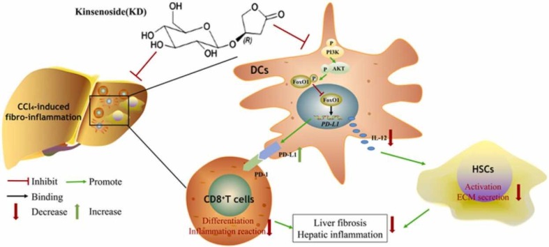
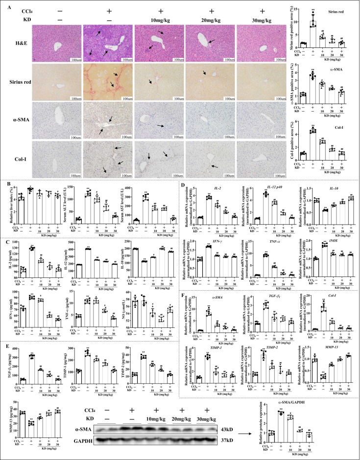
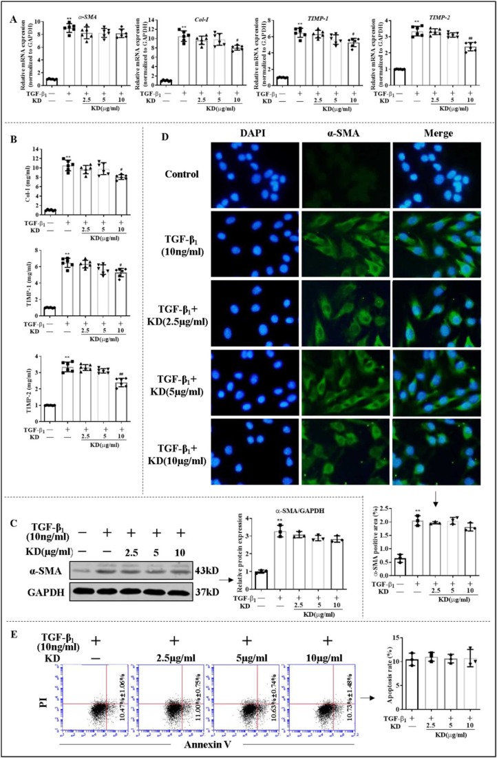
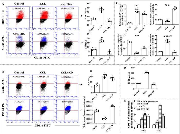
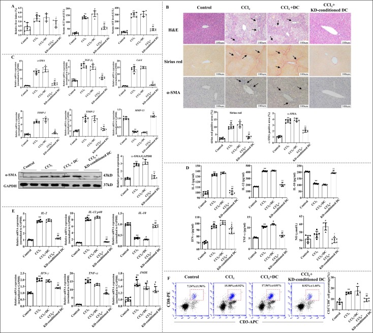
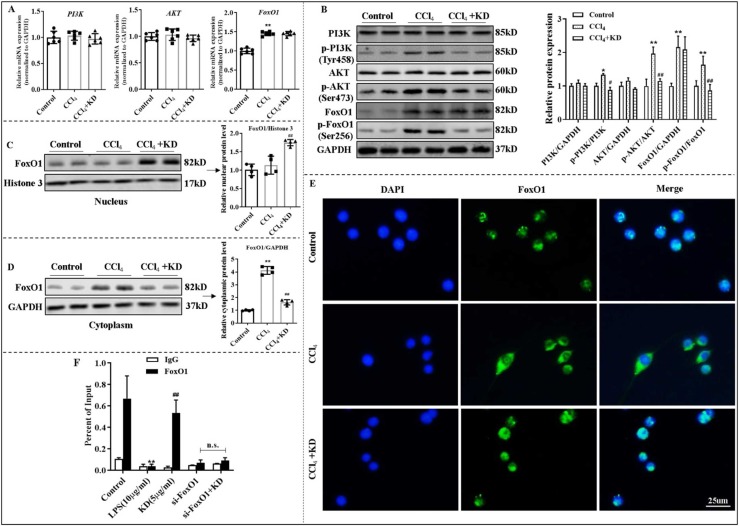
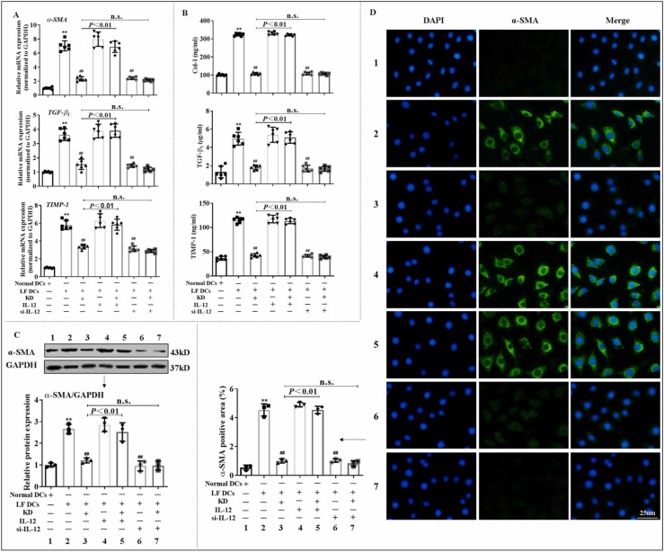
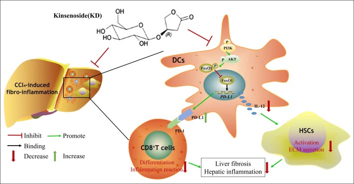
Kinsenoside (KD) exhibits anti-inflammatory and immunosuppressive effects. Dendritic cells (DCs) are critical regulators of the pathologic inflammatory milieu in liver fibrosis (LF). Herein, we explored whether and how KD repressed development of LF via DC regulation and verified the pathway involved in the process. Given our analysis, both KD and adoptive transfer of KD-conditioned DCs conspicuously reduced hepatic histopathological damage, proinflammatory cytokine release and extracellular matrix deposition in CCl4-induced LF mice. Of note, KD restrained the LF-driven rise in CD86, MHC-II, and CCR7 levels and, simultaneously, upregulated PD-L1 expression on DCs specifically, which blocked CD8+T cell activation. Additionally, KD reduced DC glycolysis, maintained DCs immature, accompanied by IL-12 decrease in DCs. Inhibiting DC function by KD disturbed the communication of DCs and HSCs with the expression or secretion of α-SMA and Col-I declined in the liver. Mechanistically, KD suppressed the phosphorylation of PI3K-AKT driven by LF or PI3K agonist, followed by enhanced nuclear transport of FoxO1 and upregulated interaction of FoxO1 with the PD-L1 promoter in DCs. PI3K inhibitor or si-IL-12 acting on DC could relieve LF, HSC activation and diminish the effect of KD. In conclusion, KD suppressed DC maturation with promoted PD-L1 expression via PI3K-AKT-FoxO1 and decreased IL-12 secretion, which blocked activation of CD8+T cells and HSCs, thereby alleviating liver injury and fibro-inflammation in LF.

Keywords: 740 Y-P (PubChem CID:90488730); Dendritic cell; Interleukin-12; Kinsenoside; Kinsenoside (PubChem CID:10422896); LY294002 (PubChem CID:3973); Liver fibro-inflammation; PI3K-AKT-FoxO1 axis; Programmed cell death ligand 1.


Conflict of interest statement

The authors declare that they have no known competing financial interests or personal relationships that could have appeared to influence the work reported in this paper.

Figures

ga1
Graphical abstract
Fig. 1
Fig. 1
KD abolished hepatic fibro-inflammation in CCl4 mice. (A) Representative images of H&E (arrows represent inflammatory infiltrations), sirius red (arrows indicate collagenous fiber deposition), and α-SMA immunohistochmical antibody (arrows represent α-SMA expression) or Col-I immunohistochmical antibody (arrows represent Col-I expression) stained liver sections. Original magnification, 200 × ; scale bar, 100 µm. (B) Levels of ALT and AST in serum, and liver index (liver wet weight/mouse body weight×100%). (C) Serum inflammatory cytokine concentrations. (D) Transcript levels of hepatic inflammatory cytokines. (E) Content of TGF-β1, TIMP-1, TIMP-2, and MMP-13 in liver homogenate and levels of hepatic α-SMA protein. (F) Hepatic mRNA levels of α-SMA, TGF-β1, Col-I, TIMP-1, TIMP-2, and MMP-13. Data expressed as mean ± SD (n = 6). *P < 0.05, **P < 0.01 relative to controls; #P < 0.05, ##P < 0.01 relative to CCl4-induced LF mice.
Fig. 2
Fig. 2
The effects of KD on HSCs (JS-1) in vitro. (A) The mRNA levels of α-SMA, Col-I, TIMP-1, and TIMP-2 in JS-1. (B) Extracellular release levels of Col-I, TIMP-1, and TIMP-2 in JS-1. (C) Levels of α-SMA protein in JS-1. (D) Immunofluorescence of α-SMA protein in JS-1. (E) Apoptotic rate of JS-1. Data expressed as mean ± SD (n = 6). **P < 0.01 relative to controls; #P < 0.05, ##P < 0.01 relative to TGF-β1 group.
Fig. 3
Fig. 3
KD inhibited the maturity and function of BMDCs ex vivo. (A) The percentage of MHC-II+ cells and CD86+ cells on CD11c+ BMDCs. (B) The percentage of CCR7 and mean fluorescence intensity of PD-L1 on CD11c+ BMDCs, as evidenced by FCM. (C) Gene levels of CCR7, PD-L1, IDO, and IL-12 p40, as quantified by qPCR. (D) Amount of IL-12 release in DC suspension, as analyzed by ELISA. (E) The DC-mediated induction of CD8+T cell proliferation, as evaluated by MLR. The ratio of CD8+T cells and DCs was 10:2 and 10:1. BMDCs were isolated from controls, CCl4-induced LF mice, and LF mice exposed to KD (30 mg/kg). Data expressed as mean ± SD (n = 6). *P < 0.05, **P < 0.01 relative to controls; #P < 0.05, ##P < 0.01 relative to CCl4 group; ΔΔP < 0.01 relative to the CD8+T lymphocytes group, which is in the absence of DC stimulation.
Fig. 4
Fig. 4
KD-conditioned DCs alleviated CCl4-induced LF and inhibited differentiation of CD8+T cells. (A) Liver index and serum ALT and AST levels. (B) KD-conditioned DCs decreased LF severity. Representative images of H&E-, sirius red-, and α-SMA histochemical antibody (brown)- stained liver sections. Original magnification, × 200; scale bar, 100 µm. (C) Transcript levels of α-SMA, TGF-β1, Col-I, TIMP-1, TIMP-2, and MMP-13 and protein levels of α-SMA in the liver. (D) Serum inflammatory cytokine levels. (E) Hepatic inflammatory cytokine transcript levels. (F) Proportion of hepatic CD8+T cells. Data expressed as mean ± SD (n = 6). P < 0.01 relative to controls; #P < 0.05, ##P < 0.01 relative to CCl4 group; ΔP < 0.05, ΔΔP < 0.01 relative to CCl4+DC group.
Fig. 5
Fig. 5
KD modulated DC function via the PI3K-AKT-FoxO1 axis in DCs. (A) Transcript levels of PI3K, AKT, and FoxO1 in DCs. (B) Total and phosphorylated levels of PI3K, AKT, and FoxO1 proteins of DCs. (C) Levels of nuclear FoxO1 protein of DCs. (D) Levels of cytoplasmic FoxO1 protein of DCs. (E) Immunofluorescence of FoxO1 (green) in DCs. BMDCs isolated from control mice or LF mice treated with or without KD (30 mg/kg). (F) ChIP assay exploring the physical binding between FoxO1 and PD-L1 promoter in DCs. IgG was used as the matched control. LPS-stimulated DCs were either exposed to KD (5 μg/mL) or not. Data expressed as mean ± SD (n = 6). *P < 0.05, **P < 0.01 relative to controls; #P < 0.05, ##P < 0.01 relative to CCl4 or LPS groups. n.s., not significant.
Fig. 6
Fig. 6
KD inhibited DC cross-priming of HSC responses via reduction of IL-12 release from DCs. (A) Transcript levels of α-SMA, TGF-β1, and TIMP-1 in JS-1, as assessed by qPCR. (B) Extracellular secretion level of Col-I, TGF-β1, and TIMP-1 in JS-1 tested by ELISA. (C-D) Protein levels of α-SMA in JS-1, as detected by WB and immunofluorescence. JS-1, with or without IL-12, was co-cultured with DCs isolated from normal or LF mice pre-treated with KD, si-IL-12, or KD+si-IL-12. Data expressed as mean ± SD (n = 6). **P < 0.01 relative to normal DCs group; ##P < 0.01 relative to LF DCs group. n.s., not significant.
Fig. 7
Fig. 7
A schematic diagram depicts the targets of KD in LF, involving PI3K-AKT-FoxO1-mediated regulation of DCs, and the interaction between DCs, CD8+ T cells, and HSCs.

References

    1. Cai X., Wang J., Wang J., Zhou Q., Yang B., He Q., Weng Q. Intercellular crosstalk of hepatic stellate cells in liver fibrosis: new insights into therapy. Pharm. Res. 2020;155 doi: 10.1016/j.phrs.2020.104720. - DOI - PubMed
    1. Connolly M.K., Bedrosian A.S., Mallen-St Clair J., Mitchell A.P., Ibrahim J., Stroud A., Pachter H.L., Bar-Sagi D., Frey A.B., Miller G. In liver fibrosis, dendritic cells govern hepatic inflammation in mice via TNF-alpha. J. Clin. Invest. 2009;119:3213–3225. doi: 10.1172/jci37581. - DOI - PMC - PubMed
    1. Weber L.W., Boll M., Stampfl A. Hepatotoxicity and mechanism of action of haloalkanes: carbon tetrachloride as a toxicological model. Crit. Rev. Toxicol. 2003;33:105–136. doi: 10.1080/713611034. - DOI - PubMed
    1. Campana L., Iredale J.P. Regression of liver fibrosis. Semin Liver Dis. 2017;37:1–10. doi: 10.1055/s-0036-1597816. - DOI - PubMed
    1. Parola M., Pinzani M. Liver fibrosis: pathophysiology, pathogenetic targets and clinical issues. Mol. Asp. Med. 2019;65:37–55. doi: 10.1016/j.mam.2018.09.002. - DOI - PubMed

Publication types