

Automated identification of pulmonary arteries and veins depicted in non-contrast chest CT scans
- PMID: 35066393
- PMCID: PMC8901546
- DOI: 10.1016/j.media.2022.102367
Automated identification of pulmonary arteries and veins depicted in non-contrast chest CT scans
Abstract
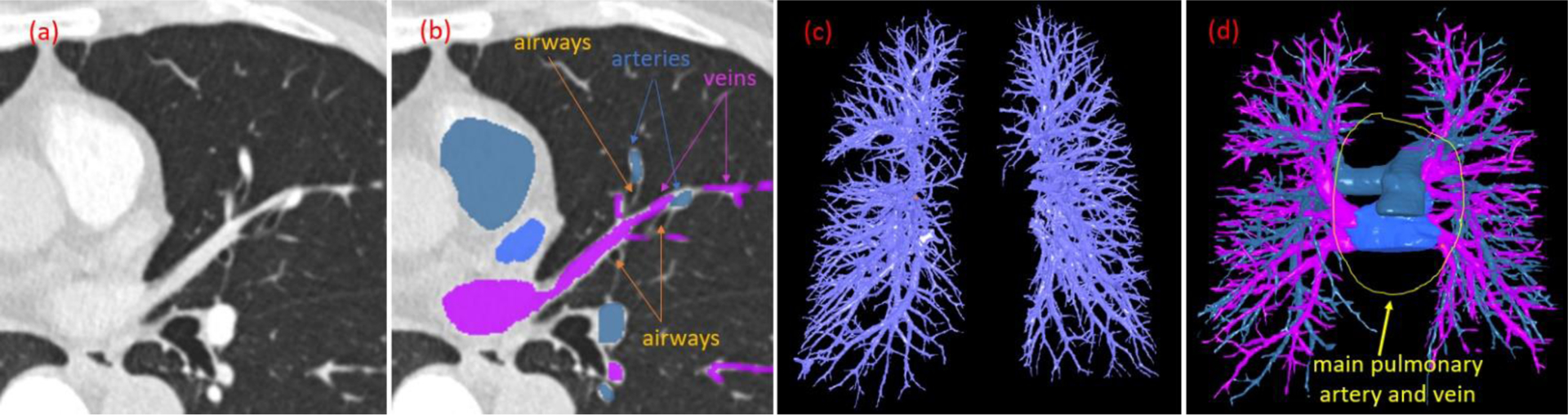
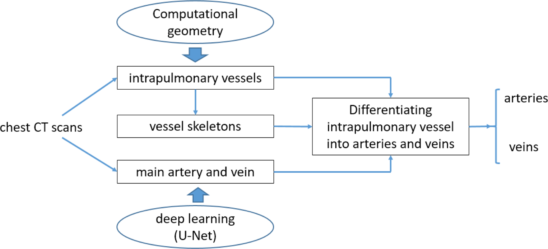
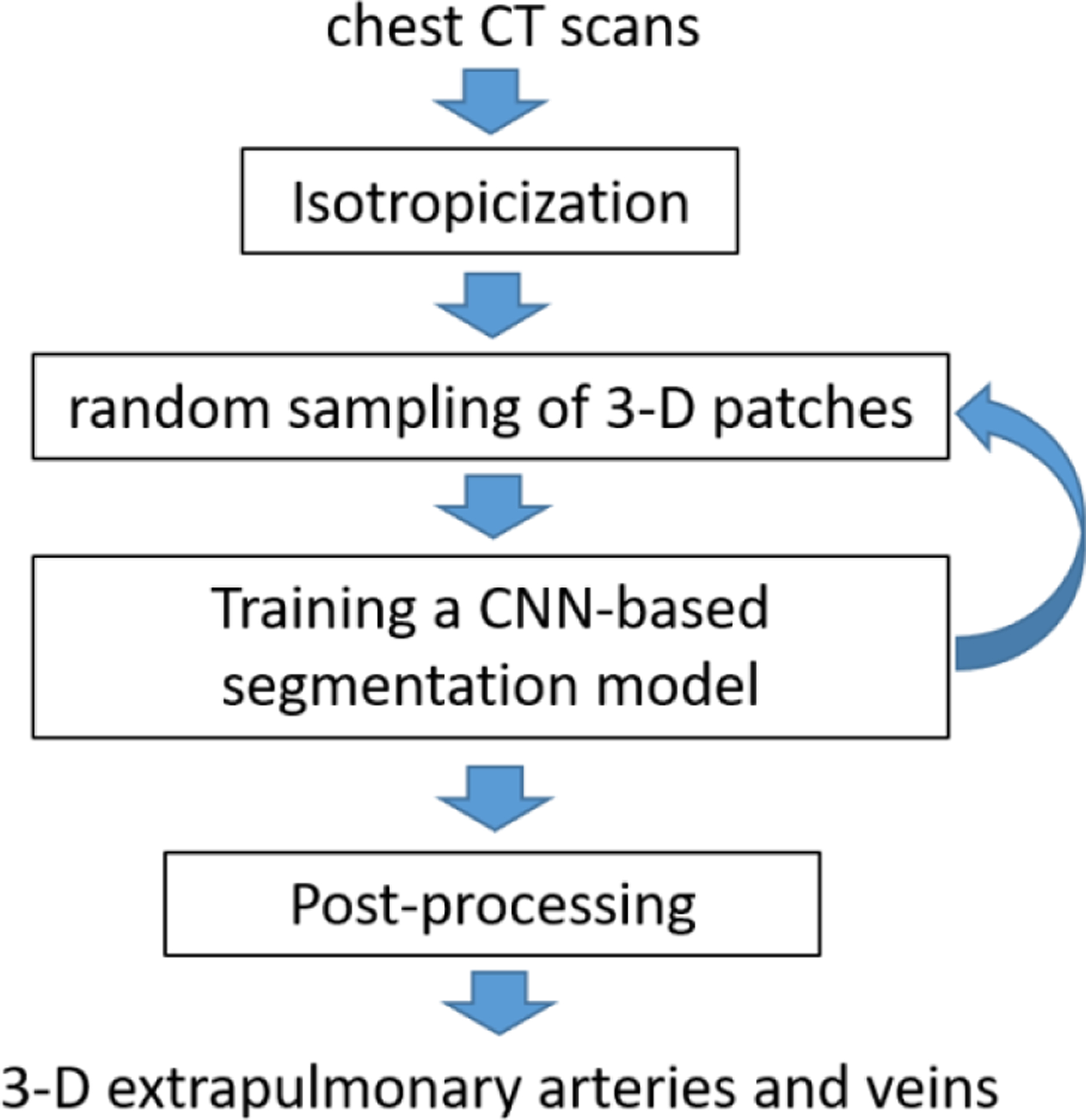
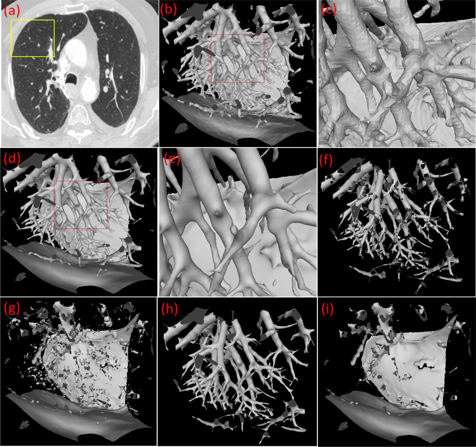
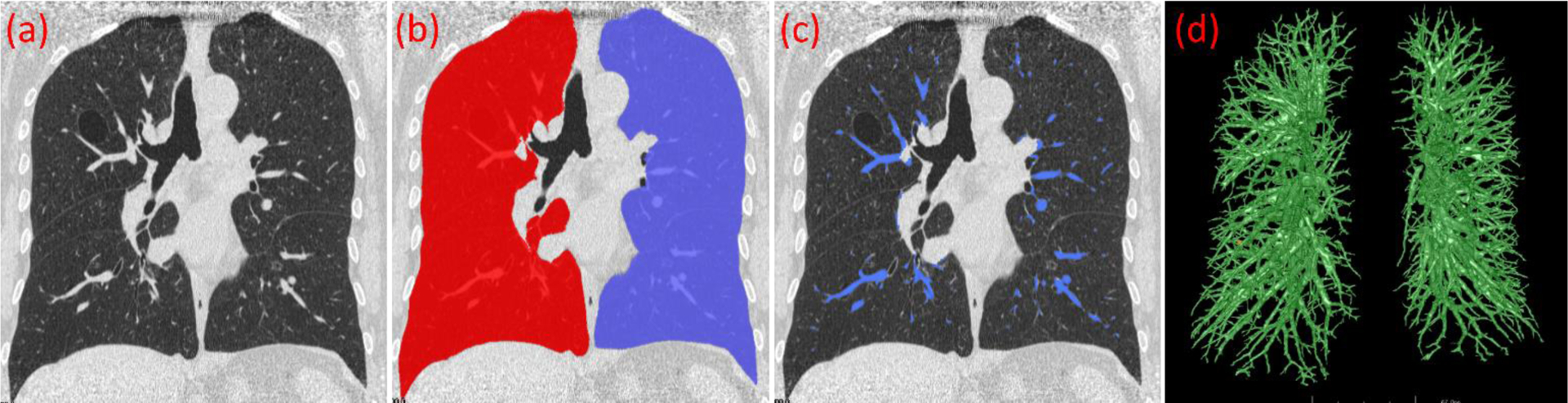
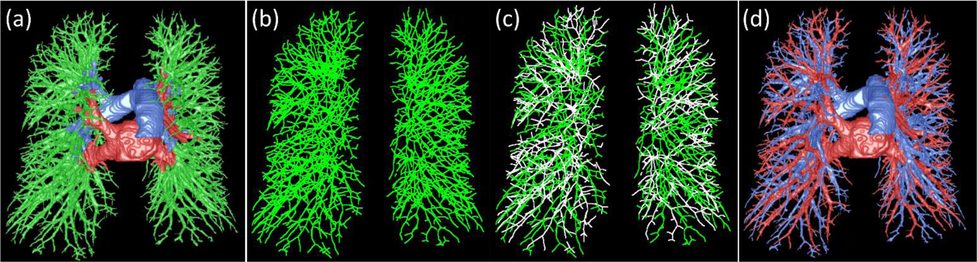
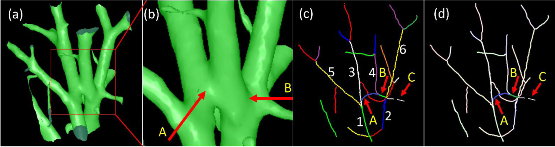
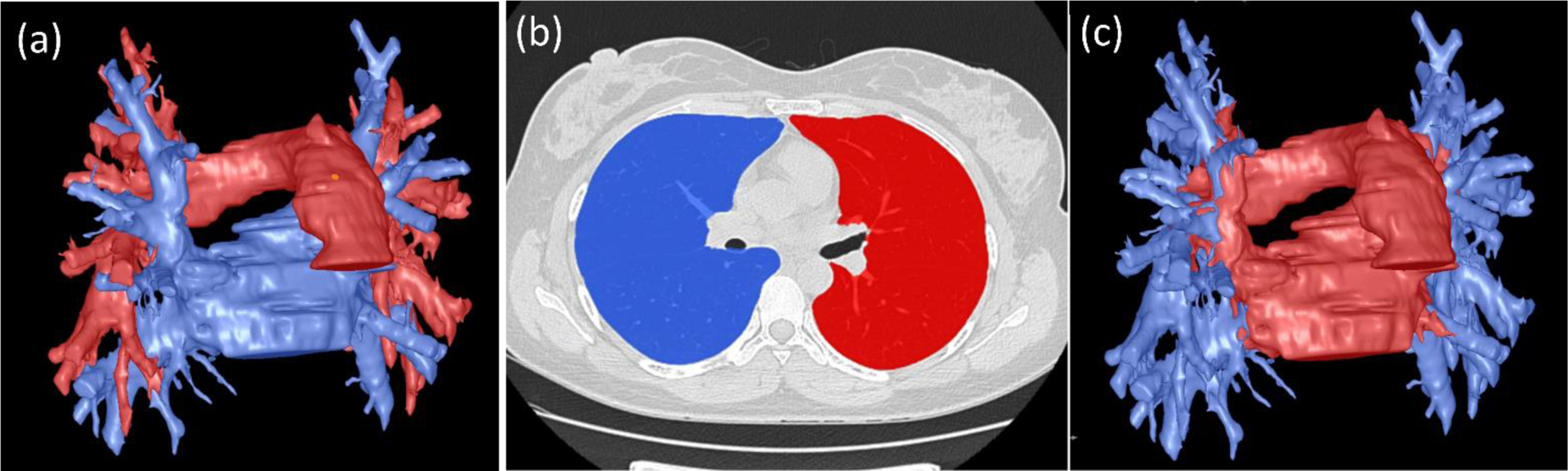
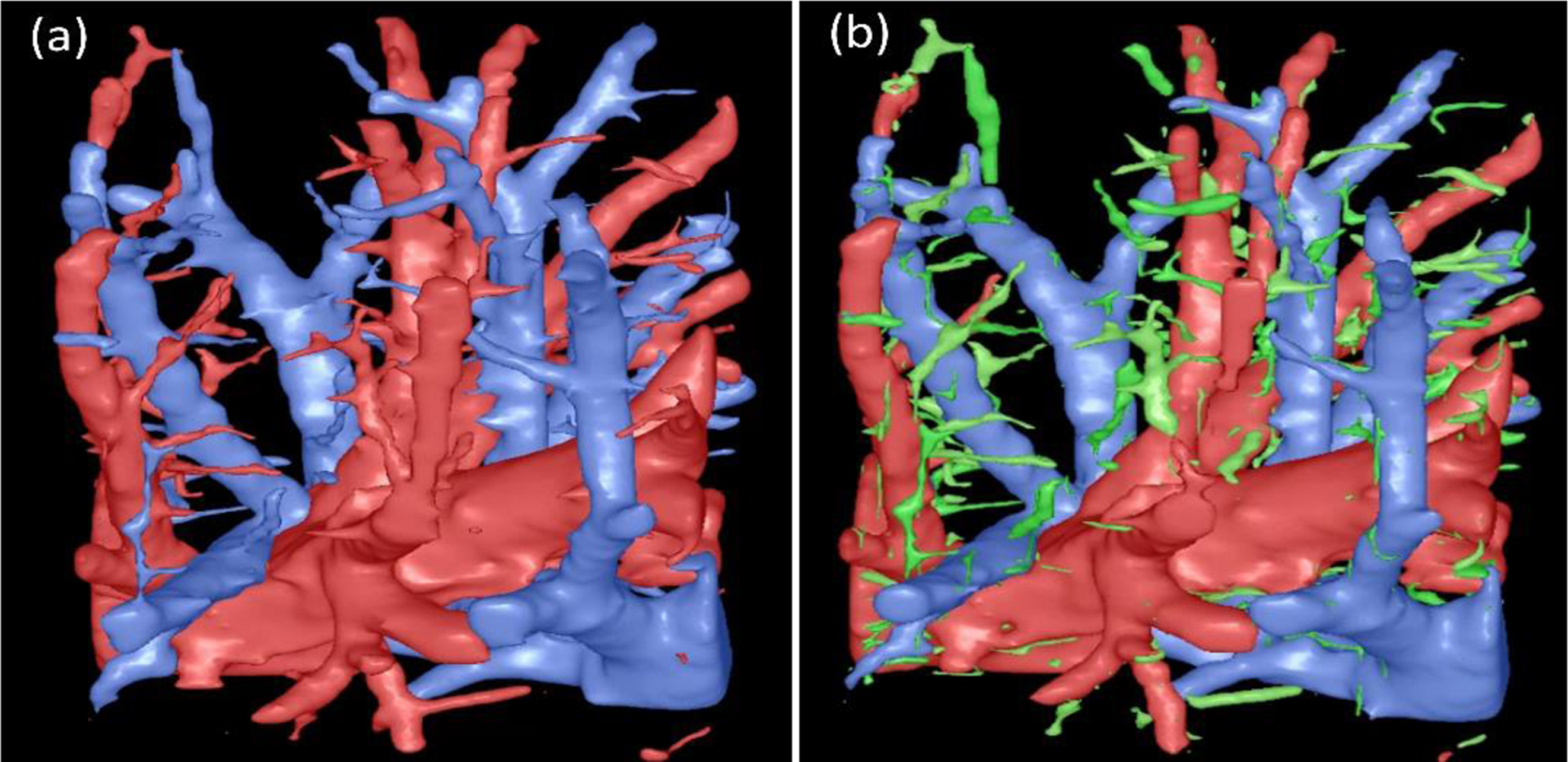
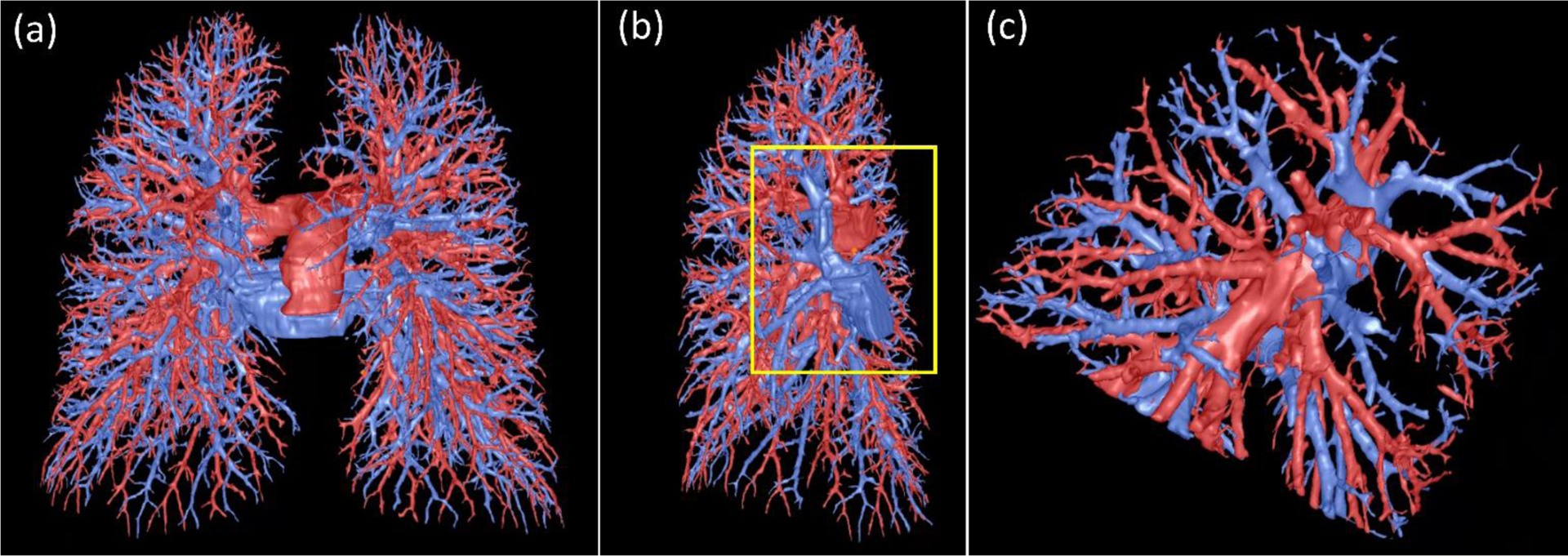
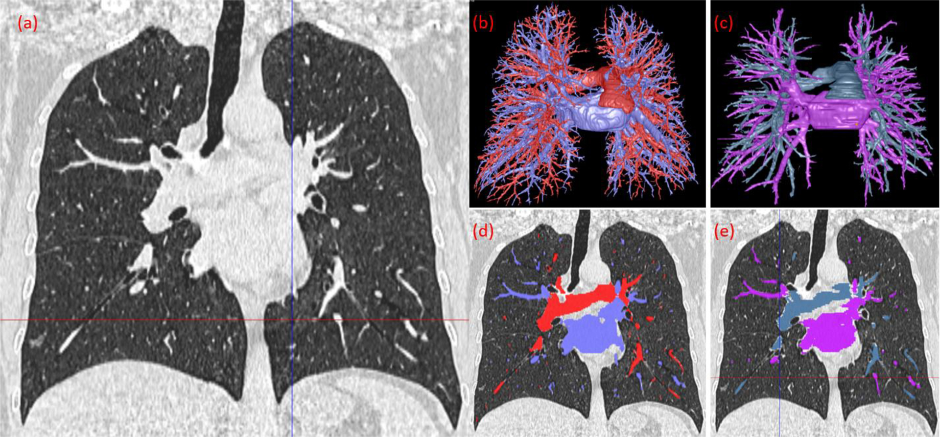
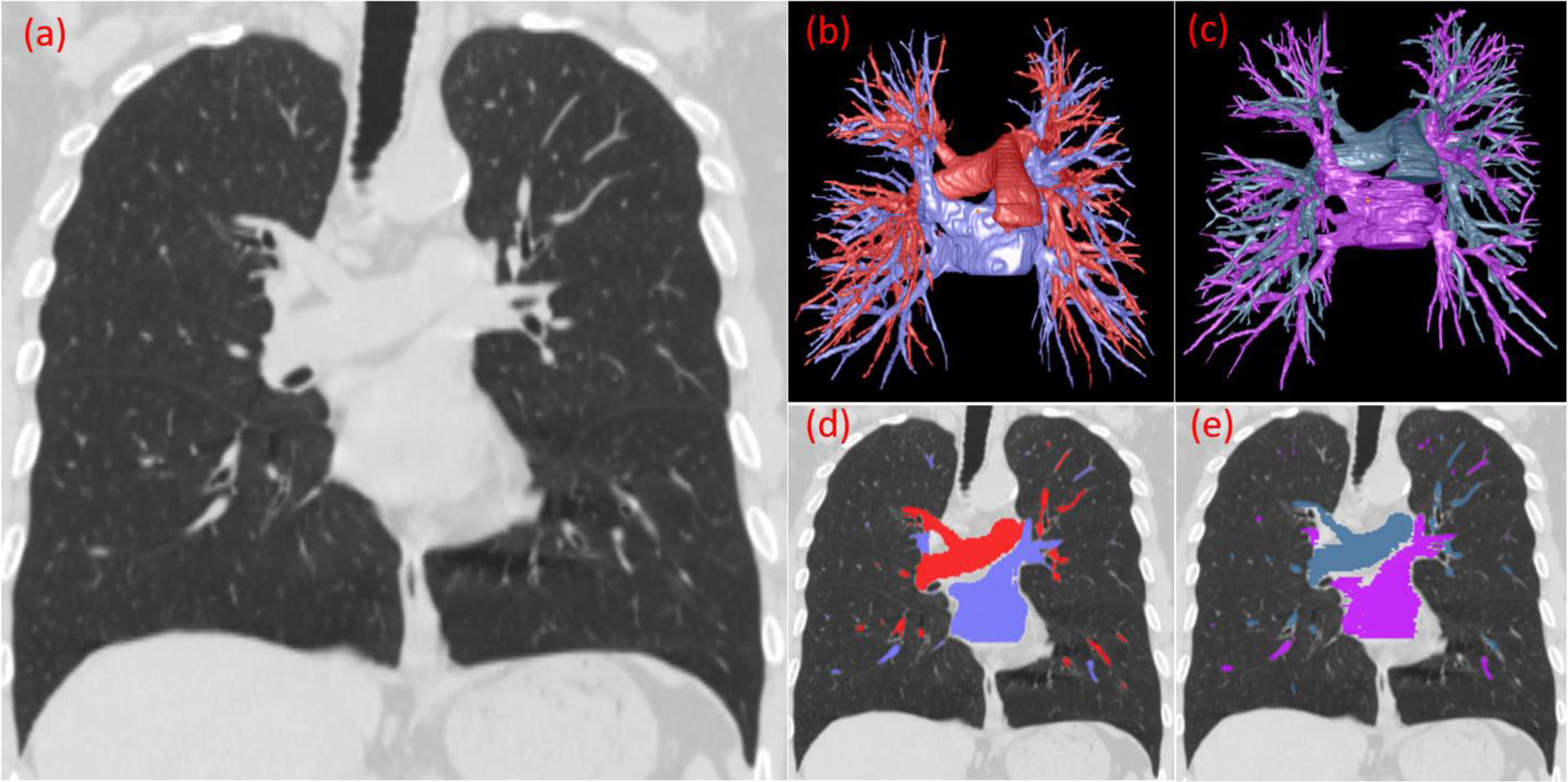
We present a novel integrative computerized solution to automatically identify and differentiate pulmonary arteries and veins depicted on chest computed tomography (CT) without iodinated contrast agents. We first identified the central extrapulmonary arteries and veins using a convolutional neural network (CNN) model. Then, a computational differential geometry method was used to automatically identify the tubular-like structures in the lungs with high densities, which we believe are the intrapulmonary vessels. Beginning with the extrapulmonary arteries and veins, we progressively traced the intrapulmonary vessels by following their skeletons and differentiated them into arteries and veins. Instead of manually labeling the numerous arteries and veins in the lungs for machine learning, this integrative strategy limits the manual effort only to the large extrapulmonary vessels. We used a dataset consisting of 120 chest CT scans acquired on different subjects using various protocols to develop, train, and test the algorithms. Our experiments on an independent test set (n = 15) showed promising performance. The computer algorithm achieved a sensitivity of ∼98% in labeling the pulmonary artery and vein branches when compared with a human expert's results, demonstrating the feasibility of our computerized solution in pulmonary artery/vein labeling.
Keywords: Artery; Computed tomography; Deep learning; Differential geometry; Vein.
Copyright © 2022 Elsevier B.V. All rights reserved.
Conflict of interest statement
Declaration of Competing Interest None.
Figures
References
-
- Ardila D, Kiraly AP, Bharadwaj S, Choi B, Reicher JJ, Peng L, Tse D, Etemadi M, Ye W, Corrado G, Naidich DP and Shetty S (2019). “End-to-end lung cancer screening with three-dimensional deep learning on low-dose chest computed tomography.” Nat Med 25(6): 954–961. - PubMed
-
- Armato SG 3rd, McLennan G, Bidaut L, McNitt-Gray MF, Meyer CR, Reeves AP, Zhao B, Aberle DR, Henschke CI, Hoffman EA, Kazerooni EA, MacMahon H, Van Beeke EJ, Yankelevitz D, Biancardi AM, Bland PH, Brown MS, Engelmann RM, Laderach GE, Max D, Pais RC, Qing DP, Roberts RY, Smith AR, Starkey A, Batrah P, Caligiuri P, Farooqi A, Gladish GW, Jude CM, Munden RF, Petkovska I, Quint LE, Schwartz LH, Sundaram B, Dodd LE, Fenimore C, Gur D, Petrick N, Freymann J, Kirby J, Hughes B, Casteele AV, Gupte S, Sallamm M, Heath MD, Kuhn MH, Dharaiya E, Burns R, Fryd DS, Salganicoff M, Anand V, Shreter U, Vastagh S and Croft BY (2011). “The Lung Image Database Consortium (LIDC) and Image Database Resource Initiative (IDRI): a completed reference database of lung nodules on CT scans.” Med Phys 38(2): 915–931. - PMC - PubMed
-
- Charbonnier JP, Brink M, Ciompi F, Scholten ET, Schaefer-Prokop CM and van Rikxoort EM (2016). “Automatic Pulmonary Artery-Vein Separation and Classification in Computed Tomography Using Tree Partitioning and Peripheral Vessel Matching.” IEEE Trans Med Imaging 35(3): 882–892. - PubMed
-
- Cornea ND, S. D, Yuan X, Balasubramanian R (2005). “Computing hierarchical curve-skeletons of 3-D objects.” Visual Comput. 21(11): 945–955.
Publication types
MeSH terms
Grants and funding
LinkOut - more resources
Full Text Sources
Other Literature Sources
Medical
