. 2022 May;257(1):109-124.
doi: 10.1002/path.5873. Epub 2022 Feb 15.

Genetic context of oncogenic drivers dictates vascular sarcoma development in aP2-Cre mice

Affiliations

Genetic context of oncogenic drivers dictates vascular sarcoma development in aP2-Cre mice

Jason A Hanna et al. J Pathol. 2022 May.

Abstract

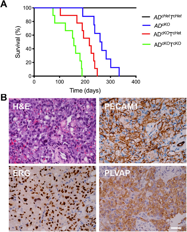
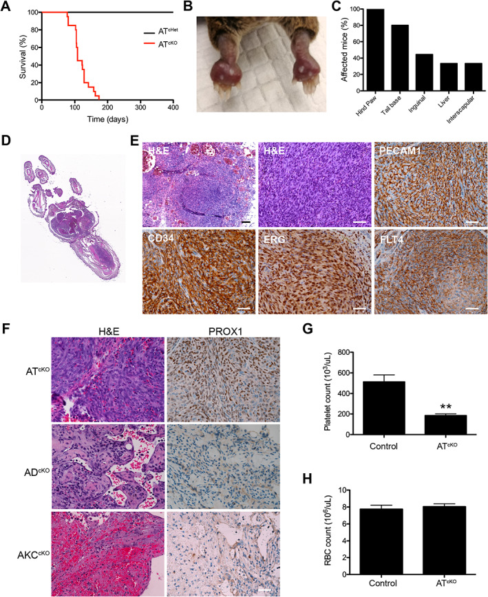
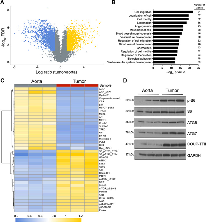
Angiosarcomas are aggressive vascular sarcomas that arise from endothelial cells and have an extremely poor prognosis. Because of the rarity of angiosarcomas, knowledge of molecular drivers and optimized treatment strategies is lacking, highlighting the need for in vivo models to study the disease. Previously, we generated genetically engineered mouse models of angiosarcoma driven by aP2-Cre-mediated biallelic loss of Dicer1 or conditional activation of KrasG12D with Cdkn2a loss that histologically and genetically resemble human tumors. In the present study, we found that DICER1 functions as a potent tumor suppressor and its deletion, in combination with either KRASG12D expression or Cdkn2a loss, is associated with angiosarcoma development. Independent of the genetic driver, the mTOR pathway was activated in all murine angiosarcoma models. Direct activation of the mTOR pathway by conditional deletion of Tsc1 with aP2-Cre resulted in tumors that resemble intermediate grade human kaposiform hemangioendotheliomas, indicating that mTOR activation was not sufficient to drive the malignant angiosarcoma phenotype. Genetic dissection of the spectrum of vascular tumors identified genes specifically regulated in the aggressive murine angiosarcomas that are also enriched in human angiosarcoma. The genetic dissection driving the transition across the malignant spectrum of endothelial sarcomas provides an opportunity to identify key determinants of the malignant phenotype, novel therapies for angiosarcoma, and novel in vivo models to further explore angiosarcoma pathogenesis. © 2022 The Authors. The Journal of Pathology published by John Wiley & Sons Ltd on behalf of The Pathological Society of Great Britain and Ireland.

Keywords: DICER1; TSC1; angiosarcoma; kaposiform hemangioendothelioma; sarcoma.


Figures

Figure 1
Figure 1
Gene expression alterations in mouse and human angiosarcomas. (A) Volcano plot of the −log10 of the FDR value versus the log fold‐change in mRNA expression in AKCcKO angiosarcomas (n = 4) versus normal wildtype aortas (n = 4) with enriched (yellow) and underrepresented (blue) with FDR < 0.05 and log2 fold‐change >1 or <−1, respectively. (B) Venn diagram of genes increased in expression with FDR < 0.05 and fold‐change >2 in ADcKO and AKCcKO (mouse angiosarcomas compared with normal aorta) and human angiosarcomas samples from previously published cohorts [9, 30]. (C) GSEA of the genes increased in expression in mouse angiosarcomas compared with the enriched human angiosarcoma gene set with a normalized enrichment score (NES) = 3.24 and FDR = 0.00. (D) Gene ontology enrichment analysis with significantly enriched biological processes (BP_FAT) in the 123 commonly enriched genes in mouse and human angiosarcomas.
Figure 2
Figure 2
KRAS activation and Cdkn2a deletion cooperate with Dicer1 loss to accelerate angiosarcoma development. (A) Kaplan–Meier survival analysis for aP2‐Cre;Dicer1 Fl/Fl (ADcKO) (blue, n = 7), aP2‐Cre;LSL‐Kras G12D ;Dicer1 Fl/Fl (AKDcKO)(green, n = 12), and aP2‐Cre;LSL‐Kras G12D ;Dicer1 Fl /+ (AKDcHet)(orange, n = 14) Log‐rank p < 0.0001. (B) Representative histology and IHC staining of an AKDcKO tumor for markers of angiosarcoma, scale bar 25 μm. (C) Kaplan–Meier survival analysis for aP2‐Cre;Cdkn2a Fl/Fl (ACcKO)(black, n = 16), aP2‐Cre;Dicer1 Fl/Fl (ADcKO)(blue, n = 7), and aP2‐Cre;Dicer1 Fl/Fl ;Cdkn2a Fl/Fl (ADcKOCcKO) (red, n = 21). Log‐rank p < 0.0001. (D) Histogram of tumor types that developed in the indicated genotype. (E) Representative histology and IHC staining for markers of angiosarcoma from an aP2‐Cre;Dicer1 F/Fl ;Cdkn2a Fl/Fl (ADcKOCcKO) tumor, scale bar 25 μm. (F) Representative histology and IHC staining for markers of histiocytic sarcomas from an aP2‐Cre;Cdkn2a Fl/Fl (ACcKO) tumor, scale bar 25 μm.
Figure 3
Figure 3
Generation of a novel murine angiosarcoma cell line and activated pathway inhibition. (A) Brightfield and tdTomato fluorescence of ADC106 cells grown in adherent conditions, scale bar 25 μm. (B) Brightfield and tdTomato fluorescence of ADC106 spheres grown in low adherence conditions, scale bar 100 μm. (C) Tube formation assay of HMEC‐1, EOMA, and ADC106 cells grown on Matrigel for 4 h, scale bar 25 μm. (D) ADC106 cell allograft formation in SCID‐Beige mice, with 1 × 106 cells injected subcutaneously in the flank (arrow). (E) Representative histology and PECAM1, ERG, and CD34 IHC staining of ADC106 cell allograft, scale bar 25 μm. (F) Growth inhibition curve of HMEC‐1 (EC50 = 19.14 nm), EOMA (EC50 = 3.5 nm), and ADC106 (EC50 = 6.3 nm) cells treated with the indicated concentration of trametinib, data presented as mean ± SEM. (G) Immunoblot analysis of cells treated with the indicated concentration of trametinib and antibodies probed indicated on the left. (H) Growth inhibition curve of HMEC‐1, EOMA, and ADC106 (EC50 = 0.96 nm) cells treated with rapamycin, data presented as mean ± SEM (*p < 0.05, **p < 0.01). (I) Immunoblot analysis of cells treated with rapamycin and antibodies probed indicated on the left.
Figure 4
Figure 4
Tsc1 deletion cooperates with Dicer1 loss accelerating tumor development. (A) Kaplan–Meier survival analysis of aP2‐Cre;Dicer1 Fl/+ ;Tsc1 Fl/+ (ADcHetTcHet)(black, n = 21), aP2‐Cre;Dicer1 Fl/Fl (ADcKO)(blue, n = 8), aP2‐Cre;Dicer1 Fl/Fl ;Tsc1 Fl/Fl (ADcKOTcKO)(green, n = 12), and aP2‐Cre;Tsc1 Fl/Fl (ATcKO) (red, n = 10) Log‐rank p < 0.0001. (B) Representative histology and IHC staining for markers of angiosarcoma in an aP2‐Cre;Dicer1 Fl/Fl ;Tsc1 Fl/Fl (ADcKOTcKO) tumor, scale bar 25 μm.
Figure 5
Figure 5
Tsc1 deletion with aP2‐Cre results in KHE‐like sarcoma tumors in the paw. (A) Kaplan–Meier tumor‐free survival analysis in aP2‐Cre;Tsc1 Fl/+ (ATcHet)(black, n = 17) and aP2‐Cre;Tsc1 Fl/Fl (ATcKO)(red, n = 20), Log‐rank p < 0.0001. (B) Representative paw tumor in an ATcKO mouse. (C) Histogram of anatomic locations of tumor development in ATcKO mice. (D) Low resolution scan of an H&E‐stained paw tumor from an ATcKO. (E) Representative histology and IHC for markers of angiosarcoma (PECAM1, CD34, ERG) and lymphatic endothelial cells (FLT4) in an ATcKO tumor, scale bar 100 μm top left (black), all others 25 μm (white). (F) Representative histology and PROX1 staining of ATcKO, ADcKO, and AKCcKO tumors, scale bar 25 μm. (G) Platelet count (**p < 0.01) and (H) RBC count in tumor‐bearing ATcKO mice (n = 4) and age‐matched control animals (n = 6), data presented as mean ± SEM.
Figure 6
Figure 6
Gene and protein expression in KHE mouse model. (A) Volcano plot of the −log10 of the FDR value versus the log ratio fold‐change in mRNA expression in aP2‐Cre;Tsc1 Fl/Fl (ATcKO) (n = 4) versus normal aorta (n = 4). Genes with FDR < 0.05 and log2 ratio > 1 (yellow) < −1 (blue). (B) Gene ontology enrichment analysis with significantly enriched biological processes (BP_FAT) in genes upregulated in tissue from ATcKO tumors compared with normal aorta with log ratio > 2 and p < 0.05 (588 genes). (C) Heatmap of the top 20 statistically significant up‐ and downregulated proteins ranked by mean difference in ATcKO tumors (n = 3) compared with normal aorta (n = 3) by RPPA. Comparisons between normal aorta and ATcKO tumors were made using the normalized linear expression and Student's t‐test with a Welch correction for unequal variance and Benjamini–Hochberg adjusted P values. (D) Immunoblot analysis of lysates from ATcKO tumors and normal aorta. Antibodies used are shown to the right.

References

    1. Dasgupta R, Fishman SJ. ISSVA classification. Semin Pediatr Surg 2014; 23: 158–161. - PubMed
    1. Putra J, Gupta A. Kaposiform haemangioendothelioma: a review with emphasis on histological differential diagnosis. Pathology 2017; 49: 356–362. - PubMed
    1. Tanas MR, Sboner A, Oliveira AM, et al. Identification of a disease‐defining gene fusion in epithelioid hemangioendothelioma. Sci Transl Med 2011; 3: 98ra82. - PubMed
    1. Florou V, Wilky BA. Current management of angiosarcoma: recent advances and lessons from the past. Curr Treat Options Oncol 2021; 22: 61. - PubMed
    1. Buehler D, Rice SR, Moody JS, et al. Angiosarcoma outcomes and prognostic factors: a 25‐year single institution experience. Am J Clin Oncol 2014; 37: 473–479. - PMC - PubMed

Publication types