. 2022;19(1):191-205.
doi: 10.1080/15476286.2021.2021677. Epub 2021 Dec 31.

Structural maturation of the HIV-1 RNA 5' untranslated region by Pr55Gag and its maturation products

Affiliations

Structural maturation of the HIV-1 RNA 5' untranslated region by Pr55Gag and its maturation products

Orian Gilmer et al. RNA Biol. 2022.

Abstract

Maturation of the HIV-1 viral particles shortly after budding is required for infectivity. During this process, the Pr55Gag precursor undergoes a cascade of proteolytic cleavages, and whilst the structural rearrangements of the viral proteins are well understood, the concomitant maturation of the genomic RNA (gRNA) structure is unexplored, despite evidence that it is required for infectivity. To get insight into this process, we systematically analysed the interactions between Pr55Gag or its maturation products (NCp15, NCp9 and NCp7) and the 5' gRNA region and their structural consequences, in vitro. We show that Pr55Gag and its maturation products mostly bind at different RNA sites and with different contributions of their two zinc knuckle domains. Importantly, these proteins have different transient and permanent effects on the RNA structure, the late NCp9 and NCp7 inducing dramatic structural rearrangements. Altogether, our results reveal the distinct contributions of the different Pr55Gag maturation products on the gRNA structural maturation.

Keywords: Gag; HIV-1; NCp15; NCp7; NCp9; Pr55Gag; RNA chaperone; RNA structure; genomic RNA; maturation.


Conflict of interest statement

The authors declare no competing interests.

Figures

Figure 1.
Figure 1.
Players and experimental strategy. (a) The Pr55Gag precursor and its nucleocapsid-containing maturation products. Scheme of the Pr55Gag, NCp15, NCp9, and NCp7 proteins are drawn in the lower part to indicate the matrix (MA), capsid (CA), spacer peptide 1 (SP1), nucleocapsid (NC), spacer peptide 2 (SP2) and p6 domains. The sequential cleavages of the Pr55Gag precursor are indicated by numbered red arrowheads and the zinc fingers located in the NC domain are indicated in green. The 3D structures of the individual domains are shown on top of the figure and these structures are artificially linked together in a linear manner as no 3D structure of the full-length Pr55Gag is available. (b) Secondary structure of the 5ʹ region of the HIV-1 genomic RNA (gRNA). One of the secondary structure models proposed in the literature is drawn to indicate the main elements present in this region, namely from 5ʹ to 3ʹ: TAR, the trans-activating region of gRNA transcription; polyA, which contains the repressed 5ʹ copy of the polyadenylation signal in its apical loop; U5:AUG (coloured in Orange), a proposed long-distance interaction between the U5 (unique in 5ʹ) region and the region surrounding the AUG initiation codon of the gag gene; PBS, the primer binding site domain to which tRNALys,3 has to be annealed to initiate reverse transcription; CU:GA (coloured in purple) a proposed long distance interaction between CU- and GA-rich regions; SL1, stem-loop 1 which contains the gRNA dimerization initiation site and is involved in gRNA packaging; SL2, which contains the main 5ʹ splice site; SL3, which is also involved in gRNA packaging. While most of these elements are present in the majority of secondary structure models proposed in the literature, the sequences forming the U5:AUG and CU:GA long distance interactions may be involved in alternative interactions. (c) Experimental strategy used in this study. gRNA 1–600 was refolded in vitro and incubated with Pr55Gag, NCp15, NCp9 or NCp7 protein (right). SHAPE was performed directly on the RNA:protein complex (Complex condition) or after treatment of the complex with AT-2 (AT-2 condition), which is able to eject Zn2+ ions from the two zinc fingers located in the NC domain. Alternatively, the protein was removed by proteinase K before performing SHAPE (ProtK condition). Controls without protein were perform for each of the three conditions (left). However, no significant differences were observed between these three controls, which were thus pooled together (NoProt condition).
Figure 2.
Figure 2.
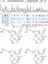
Comparison of the SHAPE reactivity profiles of the 5ʹ region of HIV-1 either alone or in complex with Pr55Gag, NCp15, NCp9, or NCp7. (a) The SHAPE reactivity profiles of nts 100–350 under NoProt, Pr55Gag Complex, NCp15 Complex, NCp9 Complex, and NCp7 Complex conditions are overlaid. (b) The significant differences between the Complex and NoProt conditions are drawn schematically and numbered. Reactivity decreases upon formation of the complexes are indicated by blue bars, while reactivity increases are indicated by red bars. Regions of decreased and increased reactivity upon complex formation are numbered in blue and red, respectively. All differences presented in this panel were statistically significant and considered to be biologically relevant (see Data analysis in the Method section). (c) These differences are plotted on the RNA secondary structure model obtained using the NoProt SHAPE reactivities as constraints, using the same colour code. Since the TAR and Poly A structures were not affected by addition of the proteins, they were omitted in the secondary structure models for clarity.
Figure 3.
Figure 3.
Effect of AT-2 on the RNA:Pr55Gag (a), RNA:NCp15 (b), RNA:NCp9 (c) and RNA:NCp7 (d) complexes. In each panel, the Complex and AT-2 SHAPE reactivity profiles are overlaid in the upper part, whereas the significant differences between the profiles are drawn in the lower part. All differences presented in the lower parts were statistically significant and considered to be biologically relevant (see Data analysis in the Method section). Reactivity increases upon AT-2 treatment are represented by blue bars, while reactivity decreases are indicated by red bars.
Figure 4.
Figure 4.
Effects of AT-2 on protected nucleotides located in SL1 (a), SL3, the AG-rich region and the junction between them (b), and PBS and SL2 regions (c). For each nt the SHAPE reactivity in the NoProt condition and in the Complex and AT-2 conditions for each of the four proteins are compared.
Figure 5.
Figure 5.
RNA chaperone activity of Pr55Gag, NCp15, NCp9, and NCp7 on the gRNA 1–600 structure. (a) The significant differences between the ProtK and NoProt SHAPE reactivity profiles are represented schematically for each protein. Reactivity increases in the ProtK conditions relative to the NoProt conditions are represented by red bars, while reactivity decreases are indicated by blue bars. All differences presented in this panel were statistically significant and considered to be biologically relevant (see Data analysis in the Method section). (b-e) Most stable secondary structure models of the 5ʹ region of HIV-1 gRNA obtained using the Pr55Gag ProtK (b), NCp15 ProtK (c), NCp9 ProtK (d), and NCp7 ProtK (e) SHAPE values as constrains. The SHAPE reactivity values are reported on the structures and colour-coded as indicated in the insert. The U5:AUG (when existing) and the CU:GA interactions are indicated in Orange and purple, respectively. Since the TAR and Poly A domains were conserved in all structures, they were omitted for clarity.
Figure 6.
Figure 6.
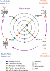
Comparison of Pr55Gag and its maturation products with respect to the contribution of the zinc fingers to RNA binding, RNA chaperone activity, transient destabilization of RNA,overall charge density, and stabilization and compaction of the gRNA dimertransient destabilization of RNA (central part) and evolution of the CU:AG interaction after exposure of the gRNA to Pr55Gag and its maturation products (outer part). Central part: Contribution of the zinc fingers to RNA binding, RNA chaperone activity, and transient destabilization of RNA were assessed semi-quantitatively from results of this study (Figs. 2, 3 & 4, and 5, respectively). Stabilization and compaction of the gRNA dimer were evaluated from published studies [18,19], and the positive charge density of each protein was calculated as the net positive charge at neutral pH divided by the number of amino acids in the protein. The positive charge density is 0.042, 0.107, 0.225 and 0.236 for Pr55Gag, NCp15, NCp9, and NCp7, respectively. Outer part: The CU:AG interaction resulting from exposure to NCp9 and NCp7 prevents formation of the U5:AUG interaction.

References

    1. Swanstrom R, Wills JW.. Synthesis, assembly, and processing of viral proteins [Internet]. In: Coffin JM, Hughes SH, and Varmus HE, editors. Retroviruses. Cold Spring Harbor (NY):Cold Spring Harbor Laboratory Press; 1997. Available from: http://www.ncbi.nlm.nih.gov/books/NBK19456/ - PubMed
    1. Sundquist WI, Kräusslich H-G. HIV-1 assembly, budding, and maturation. Cold Spring Harb Perspect Med. 2012;2:a006924. - PMC - PubMed
    1. Freed EO. HIV-1 assembly, release and maturation. Nat Rev Microbiol. 2015;13:484–496. - PMC - PubMed
    1. Kleinpeter AB, Freed EO. HIV-1 maturation: lessons learned from inhibitors. Viruses. 2020;12(9):940. 10.3390/v12090940 - DOI - PMC - PubMed
    1. Fu W, Gorelick RJ, Rein A. Characterization of human immunodeficiency virus type 1 dimeric RNA from wild-type and protease-defective virions. J Virol. 1994;68:5013–5018. - PMC - PubMed

Publication types

MeSH terms

Substances