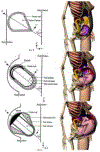
Radiation Absorbed Dose to the Embryo and Fetus from Radiopharmaceuticals
- PMID: 35067360
- PMCID: PMC8923960
- DOI: 10.1053/j.semnuclmed.2021.12.007
Radiation Absorbed Dose to the Embryo and Fetus from Radiopharmaceuticals
Abstract
Nuclear medicine procedures are generally avoided during pregnancy out of concern for the radiation dose to the fetus. However, for clinical reasons, radiopharmaceuticals must occasionally be administered to pregnant women. The procedures most likely to be performed voluntarily during pregnancy are lung scans to diagnose pulmonary embolism and 18F-fluoro-2-deoxyglucose (18F-FDG) scans for the staging of cancers. This article focuses on the challenges of fetal dose calculation after administering radiopharmaceuticals to pregnant women. In particular, estimation of the fetal dose is hampered by the lack of fetal biokinetic data of good quality and is subject to the variability associated with methodological choices in dose calculations, such as the use of various anthropomorphic phantoms and modeling of the maternal bladder. Despite these sources of uncertainty, the fetal dose can be reasonably calculated within a range that is able to inform clinical decisions. Current dose estimates suggest that clinically justified nuclear medicine procedures should be performed even during pregnancy because the clinical benefits for the mother and the fetus outweigh the small and purely hypothetical radiation risk to the fetus. In addition, the fetal radiation dose should be minimized without compromising image quality, such as by encouraging bladder voiding and by using positron emission tomography (PET)/magnetic resonance imaging (MRI) devices or high-sensitivity PET scanners that generate images of good quality with a lower injected activity.
Copyright © 2021. Published by Elsevier Inc.
Figures


Similar articles
-
18F-FDG Fetal Dosimetry Calculated with PET/MRI.J Nucl Med. 2022 Oct;63(10):1592-1597. doi: 10.2967/jnumed.121.263561. Epub 2022 Feb 3. J Nucl Med. 2022. PMID: 35115366 Free PMC article.
-
Fetal and maternal absorbed dose estimates for positron-emitting molecular imaging probes.J Nucl Med. 2014 Sep;55(9):1459-66. doi: 10.2967/jnumed.114.141309. Epub 2014 Jul 14. J Nucl Med. 2014. PMID: 25024424
-
New Fetal Dose Estimates from 18F-FDG Administered During Pregnancy: Standardization of Dose Calculations and Estimations with Voxel-Based Anthropomorphic Phantoms.J Nucl Med. 2016 Nov;57(11):1760-1763. doi: 10.2967/jnumed.116.173294. Epub 2016 Jun 3. J Nucl Med. 2016. PMID: 27261522
-
Administered radionuclides in pregnancy.Teratology. 1999 Apr;59(4):236-9. doi: 10.1002/(SICI)1096-9926(199904)59:4<236::AID-TERA9>3.0.CO;2-6. Teratology. 1999. PMID: 10331526 Review.
-
The lung cancers: staging and response, CT, 18F-FDG PET/CT, MRI, DWI: review and new perspectives.Br J Radiol. 2023 Aug;96(1148):20220339. doi: 10.1259/bjr.20220339. Epub 2023 May 17. Br J Radiol. 2023. PMID: 37097296 Free PMC article. Review.
Cited by
-
EANM practice guidelines for an appropriate use of PET and SPECT for patients with epilepsy.Eur J Nucl Med Mol Imaging. 2024 Jun;51(7):1891-1908. doi: 10.1007/s00259-024-06656-3. Epub 2024 Feb 23. Eur J Nucl Med Mol Imaging. 2024. PMID: 38393374 Free PMC article.
-
Medicolegal considerations associated with cancer during pregnancy.Abdom Radiol (NY). 2023 May;48(5):1637-1644. doi: 10.1007/s00261-022-03776-y. Epub 2022 Dec 20. Abdom Radiol (NY). 2023. PMID: 36538081 Review.
-
18F-FDG Fetal Dosimetry Calculated with PET/MRI.J Nucl Med. 2022 Oct;63(10):1592-1597. doi: 10.2967/jnumed.121.263561. Epub 2022 Feb 3. J Nucl Med. 2022. PMID: 35115366 Free PMC article.
References
-
- Grigoryan A, Bouyoucef S, Sathekge M, et al. Development of nuclear medicine in Africa. Clinical and translational imaging. 2021.
-
- IMV 2020. PET Imaging Market Summary Report.
-
- Shao F, Chen Y, Huang Z, Cai L, Zhang Y. Unexpected Pregnancy Revealed on 18F-NaF PET/CT. Clin Nucl Med. 2016;41:e202–203. - PubMed
-
- Brent RL. Saving lives and changing family histories: appropriate counseling of pregnant women and men and women of reproductive age, concerning the risk of diagnostic radiation exposures during and before pregnancy. Am J Obstet Gynecol. 2009;200:4–24. - PubMed
Publication types
MeSH terms
Substances
Grants and funding
LinkOut - more resources
Full Text Sources

