. 2022 Mar 3;84(3):325-329.
doi: 10.1292/jvms.21-0631. Epub 2022 Jan 21.

Rapid determination of pathogens in mastitic milk of dairy cows using Gram staining

Affiliations

Rapid determination of pathogens in mastitic milk of dairy cows using Gram staining

Naoki Suzuki et al. J Vet Med Sci. .

Abstract

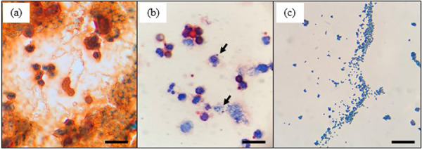
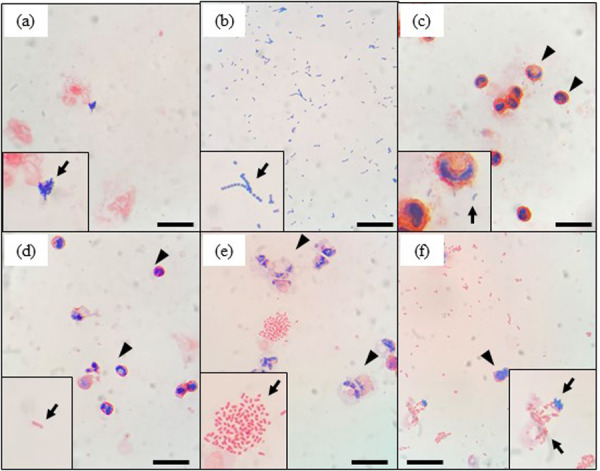
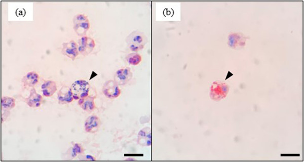
This study aimed to determine whether causative pathogens in mastitic milk can be determined by Gram staining after the centrifugation of milk. Gram staining was performed using unconcentrated and concentrated milk cells. Using this method, we found that the background of microscopic image of unconcentrated milk cells was complex and bacteria were difficult to detect. In contrast, the background of the smears in the concentrated milk cells was translucent, and bacterial and somatic cells were clearly visible. The sensitivity and specificity of the Gram staining of concentrated milk cells were 84.4% and 86.0% and 50.0% and 94.5% for the detection of gram-positive and gram-negative bacteria, respectively. The presented method provides a simple and inexpensive means of determining mastitis-causing pathogens.

Keywords: Gram staining; dairy cow; diagnosis; mastitis.


Conflict of interest statement

The authors declare that they have no conflicts of interest associated with this study.

Figures

Fig. 1.
Fig. 1.
Microscopic images of gram-stained unconcentrated milk cells without centrifugation (a), concentrated milk cells with centrifugation (b), and isolates from the same mastitic milk sample (c). Arrows in (b) indicate the causal mastitis bacteria in milk. Scale bars=20 μm.
Fig. 2.
Fig. 2.
Microscopic images of gram-stained mastitic milk samples after centrifugation. For each image, insets show enlarged images of bacteria. The arrows in each image indicate a representative image of bacteria. Clustered gram-positive cocci (a and f), chains of gram-positive cocci (b), gram-positive bacilli (c), and gram-negative bacilli (d, e, and f) were observed. Arrowheads in d, e, and f indicate leukocytes in milk. Scale bars=20 μm.
Fig. 3.
Fig. 3.
Microscopic images showing leukocyte phagocytosis of gram-positive cocci (a, arrowhead) and gram-negative bacilli (b, arrowhead). Scale bars=10 μm.

References

    1. Ashraf A., Imran M.2018. Diagnosis of bovine mastitis: from laboratory to farm. Trop. Anim. Health Prod. 50: 1193–1202. doi: 10.1007/s11250-018-1629-0 - DOI - PubMed
    1. Breed R. S., Brew J. D.1917. The control of public milk supplies by the use of the microscopic method. J. Dairy Sci. 1: 259–271. doi: 10.3168/jds.S0022-0302(17)94379-6 - DOI
    1. Britten A. M.2012. The role of diagnostic microbiology in mastitis control programs. Vet. Clin. North Am. Food Anim. Pract. 28: 187–202. doi: 10.1016/j.cvfa.2012.03.006 - DOI - PubMed
    1. Farnsworth R. J., Sorensen D. K.1972. Prevalence and species distribution of yeast in mammary glands of dairy cows in Minnesota. Can. J. Comp. Med. 36: 329–332. - PMC - PubMed
    1. Fuenzalida M. J., Ruegg P. L.2019. Negatively controlled, randomized clinical trial to evaluate intramammary treatment of nonsevere, gram-negative clinical mastitis. J. Dairy Sci. 102: 5438–5457. doi: 10.3168/jds.2018-16156 - DOI - PubMed

Substances