

A Rare Cause of Left Shoulder Pain in a Peritoneal Dialysis Patient
- PMID: 35067671
- PMCID: PMC8800465
- DOI: 10.12659/AJCR.933223
A Rare Cause of Left Shoulder Pain in a Peritoneal Dialysis Patient
Abstract
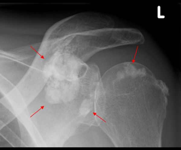
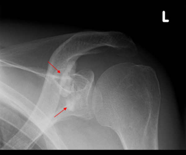
BACKGROUND Non-specific pain of connective tissues and joints is one of the most frequently expressed patient concerns in everyday practice. The most common cause is osteo-degenerative changes in the cartilage and/or joint system. Metastatic calcification is a rare and initially often overlooked cause of persistent, therapy-resistant pain of connective tissues and joint apparatus in end-stage renal disease (ESRD) patients on dialysis therapy. These calcifications are induced by persistent hyperphosphatemia/hyperparathyroidism and can occur in various organs, including joints, tendons, heart valves, soft tissues, and blood vessels. CASE REPORT We report on a 46-year-old male patient with ESRD due to cANCA-associated systemic vasculitis. The patient evolved unfavorably to end-stage renal failure and started continuous ambulatory peritoneal dialysis (CAPD). Four years after initiation of CAPD, the patient reported having painful motion of the left shoulder, and symptomatic physiotherapy and non-steroidal-anti-inflammatory-drugs (NSAIDs) were prescribed. An X-ray examination of the left shoulder showed severe periarticular calcifications. Repeated nutritional counselling was offered, and intensive phosphate-binder therapy was administered, resulting in a reduction in phosphate levels from 2.10 mmol at the time of diagnosis to 1.26 mmol/l 16 months later. Radiological reevaluation showed a near complete resolution of the periarticular calcifications. CONCLUSIONS Metastatic calcifications may arise in ESRD patients despite only moderately elevated blood phosphate levels. Intensive measures to reduce the phosphate load to normal levels should be implemented and can lead to almost complete resolution of ectopic calcifications in affected patients.
Conflict of interest statement
Figures
References
-
- Labidi J, Ben Ariba Y, Ben Gabsia A, et al. Severe metastatic calcifications in a hemodialysis patient. Saudi J Kidney Dis Transpl. 2016;27:1037–42. - PubMed
-
- Agarwal R, Burns RR, Vergne-Marini P. Paraparesis due to massive ectopic paravertebral calcification in a patient on maintenance hemodialysis. Am J Kidney Dis. 1993;22:717–20. - PubMed
-
- Cowlam TE, Bucknall TE. Cutaneous ectopic breast calcification in a haemodialysis patient. Breast. 2003;12:342–44. - PubMed
-
- Elghobashy M, Vaquas S, Elshafie M, et al. Unusual presentation of mam-mary calciphylaxis in a patient on long-standing renal dialysis. Pathobiology. 2020;87:317–21. - PubMed
Publication types
MeSH terms
LinkOut - more resources
Full Text Sources
