Review
. 2022 Mar;306(1):58-75.
doi: 10.1111/imr.13066. Epub 2022 Jan 23.

Immune dynamics in the CNS and its barriers during homeostasis and disease

Affiliations
Review

Immune dynamics in the CNS and its barriers during homeostasis and disease

Monica W Buckley et al. Immunol Rev. 2022 Mar.

Abstract

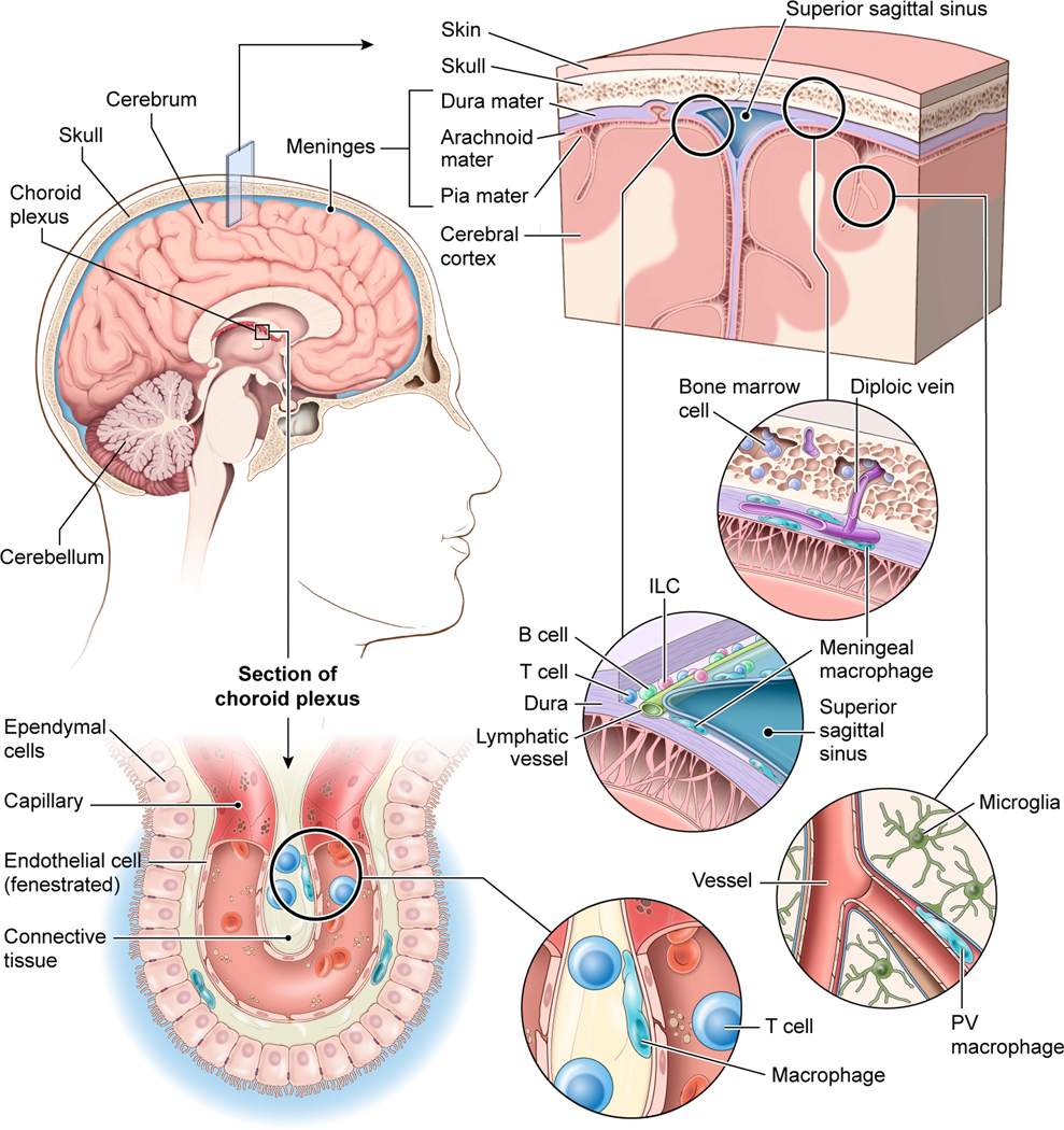
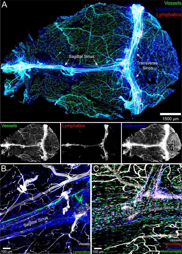
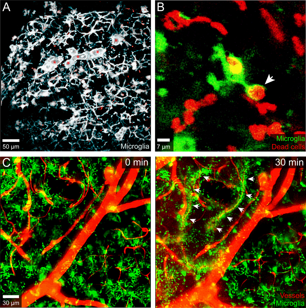
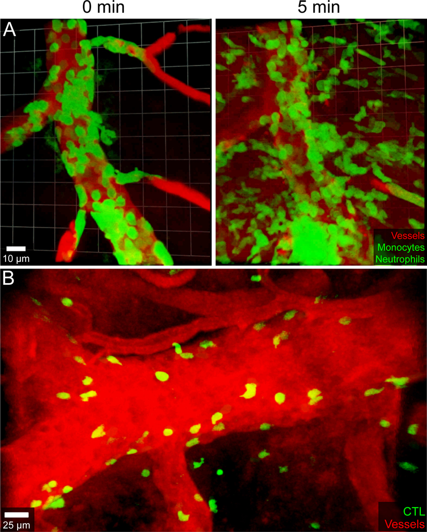
The central nervous system (CNS) has historically been viewed as an immunologically privileged site, but recent studies have uncovered a vast landscape of immune cells that reside primarily along its borders. While microglia are largely responsible for surveying the parenchyma, CNS barrier sites are inhabited by a plethora of different innate and adaptive immune cells that participate in everything from the defense against microbes to the maintenance of neural function. Static and dynamic imaging studies have revolutionized the field of neuroimmunology by providing detailed maps of CNS immune cells as well as information about how these cells move, organize, and interact during steady-state and inflammatory conditions. These studies have also redefined our understanding of neural-immune interactions at a cellular level and reshaped our conceptual view of immune privilege in this specialized compartment. This review will focus on insights gained using imaging techniques in the field of neuroimmunology, with an emphasis on anatomy and CNS immune dynamics during homeostasis, infectious diseases, injuries, and aging.

Keywords: Alzheimer's; brain; dura; glymphatics; infection; intravital; lymphatics; meninges; microglia; monocytes; neuroimmunology; neutrophils; sinuses; stroke; traumatic brain injury; two-photon microscopy; vasculature; virus.


Conflict of interest statement

Conflicts of Interests

The authors have no conflicts of interest to report.

Figures

Figure 1.
Figure 1.. The immune defense of different CNS barriers.
The brain is confined by the skull bone which contains pockets of bone marrow connected to the dura mater via diploic veins. The dura mater is vascularized by vessels that resemble those found in the periphery (i.e., they do not have tight junctions). These ‘open’ vessels are lined by meningeal macrophages. The dura mater also has large venous drains referred to as sinuses. These fenestrated sinuses are lined by lymphatic vessels as well as a diverse collection of immune cells, including macrophages, T cells, B cells, and innate lymphoid cells (ILCs), among others. Beneath the dura mater are the other two meningeal layers – the arachnoid mater and pia mater – which together are referred to as the leptomeninges. Blood vessels within the leptomeninges and brain parenchyma are sealed by tight junctions, which represents an important CNS barrier. Perivascular (PV) macrophages reside in the spaces between blood vessels that enter the brain and astrocytic foot processes comprising the blood brain barrier. The brain parenchyma itself is surveilled by microglia. The choroid plexus is another CNS barrier responsible for producing cerebral spinal fluid. It contains fenestrated vessels that reside behind ependymal cells connected by tight junctions. The choroid plexus is protected by macrophages and T cells as well as other immune cells.
Figure 2.
Figure 2.. Anatomy of the meninges during steady state.
A.) The naïve meninges beneath the skull bone of an 8-week-old C57BL/6J mouse were harvested and imaged in 3D using confocal microscopy. The whole mount consisting primarily of dura and arachnoid mater shows the distribution of blood vessels labeled intravenously with fluorescent tomato lectin (green), CD45+ leukocytes (blue), and Lyve1+ lymphatic vessels (red). The individual grayscale images for each channel are shown beneath the 3-color overlay. The sagittal and transverse sinuses are also labeled. Note that the meningeal lymphatics are juxtaposed to the dural venous sinuses. B.) A three color zoomed confocal image from a meningeal whole mount shows a Lyve1+ lymphatic vessel adjacent to the superior sagittal sinus. CD45+ leukocytes are shown in blue and tomato lectin+ blood vessels in white. C.) A confocal image captured from a meningeal whole mount shows the distribution of CCR2rfp/+ monocytes (red) and CX3CR1gfp/+ meningeal macrophages (green) along the superior sagittal sinus. Blood vessels are shown in white.
Figure 3.
Figure 3.. Microglia responses to mTBI and cerebrovascular injury.
A.) Intravital two-photon microscopy was used to image the mTBI response in a CX3CR1gfp/+ CCR2rfp/+ mouse immediately following a focal meningeal compression injury. A 3D time lapse was captured through a 20μm thinned skull window for the first hour after injury. This maximal projection image at 30 min post-injury shows the microglial response along the glia limitans superficialis. Most of the microglia (white) in the image are forming honeycomb-like structures designed to seal the gaps between individual surface associated astrocytes (not labeled). The amoeboid microglia denoted with red asterisks and resembling ‘jellyfish’ are responsible for clearing debris. B.) A CX3CR1gfp/+ microglia (green) captured in another two-photon time lapse after mTBI is shown acquiring a dead cell (red) labeled transcranially with propidium iodide. The microglial process acquiring the dead cell is denoted with a white arrowhead. C.) Two-imaging was used to capture a time lapse through the thinned skull window of CX3CR1gfp/+ mouse immediately after inducing a cerebrovascular injury in the neocortex with transcranial ultrasound + intravenous microbubbles. Two time points are shown in the time lapse: 0- and 30-minutes post-injury. The microglia are naïve at time point zero but then extend processes and envelop individual damaged blood vessels forming rosettes by 30 min (denoted with white arrowheads). Blood vessels (red) were visualized by injecting Evans Blue intravenously.
Figure 4.
Figure 4.. Immune mediated damage to CNS vasculature during viral meningitis and cerebral malaria.
A.) An intravital two-photon time lapse captured through the thinned skull window of a LysMgfp/+ mouse six days following intracerebral inoculation of LCMV Armstrong shows myelomonocytic cells (green) synchronously extravasating from a meningeal blood vessel. Quantum dots (red) were injected intravenously to visualize blood vessels. Note the massive extravasation of myelomonocytic cells at 5 min, which coincides with leakage of quantum dots into the extravascular space. B.) Parasite-specific cytotoxic lymphocytes (CTL, green) tagged with a fluorescent protein were visualized at the peak of disease (day 6) in a C57BL/6J mouse infected intravenously with 106 red blood cells parasitized by Plasmodium berghei ANKA. CTL were observed attacking meningeal and cerebral vasculature, arresting along both the luminal and abluminal surfaces. These CTL engagements are responsible for fatal vascular breakdown during cerebral malaria in both mice and humans. Blood vessels were visualized with Evans Blue (red).

References

    1. Engelhardt B, Vajkoczy P, Weller RO. The movers and shapers in immune privilege of the CNS. Nat Immunol. 2017;18(2):123–131. - PubMed
    1. Louveau A, Harris TH, Kipnis J. Revisiting the Mechanisms of CNS Immune Privilege. Trends Immunol. 2015;36(10):569–577. - PMC - PubMed
    1. MEDAWAR PB. Immunity to homologous grafted skin; the fate of skin homografts transplanted to the brain, to subcutaneous tissue, and to the anterior chamber of the eye. Br J Exp Pathol. 1948;29(1):58–69. - PMC - PubMed
    1. Forrester JV, McMenamin PG, Dando SJ. CNS infection and immune privilege. Nat Rev Neurosci. 2018;19(11):655–671. - PubMed
    1. Engelhardt B, Coisne C. Fluids and barriers of the CNS establish immune privilege by confining immune surveillance to a two-walled castle moat surrounding the CNS castle. Fluids Barriers CNS. 2011;8(1):4. - PMC - PubMed

Publication types