Review
. 2022 Jan 7:15:787164.
doi: 10.3389/fncir.2021.787164. eCollection 2021.

Genetic Mechanisms Underlying the Evolution of Connectivity in the Human Cortex

Affiliations
Review

Genetic Mechanisms Underlying the Evolution of Connectivity in the Human Cortex

Ewoud R E Schmidt et al. Front Neural Circuits. .

Abstract

One of the most salient features defining modern humans is our remarkable cognitive capacity, which is unrivaled by any other species. Although we still lack a complete understanding of how the human brain gives rise to these unique abilities, the past several decades have witnessed significant progress in uncovering some of the genetic, cellular, and molecular mechanisms shaping the development and function of the human brain. These features include an expansion of brain size and in particular cortical expansion, distinct physiological properties of human neurons, and modified synaptic development. Together they specify the human brain as a large primate brain with a unique underlying neuronal circuit architecture. Here, we review some of the known human-specific features of neuronal connectivity, and we outline how novel insights into the human genome led to the identification of human-specific genetic modifiers that played a role in the evolution of human brain development and function. Novel experimental paradigms are starting to provide a framework for understanding how the emergence of these human-specific genomic innovations shaped the structure and function of neuronal circuits in the human brain.

Keywords: dendritic morphology; evolution; human brain; human neuron physiology; human-specific genes; neuronal connectivity; synapses.


Conflict of interest statement

The authors declare that the research was conducted in the absence of any commercial or financial relationships that could be construed as a potential conflict of interest.

Figures
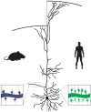
Figure 1
Figure 1
Evolution of pyramidal neuron morphology. In human pyramidal neurons (PNs), a larger number of branch points leads to a more extensive and complex dendritic tree. Each branch also contains a larger number of synapses, resulting in an overall increase in the number of synapses made onto human PNs. In addition, because of the increased cortical thickness of the human brain, the apical dendrites of human PNs extend over a longer distance to reach layer I.
Figure 2
Figure 2
Functional properties of human pyramidal neurons. The length of the apical dendrite of human pyramidal neurons (PNs) can lead to electrical isolation of the distal dendritic domain. In deep layer cortical neurons this results in disrupted coupling between the soma and the distal tuft. The summation of activity in multiple dendritic segments, through non-linear integration rules, are likely required to couple distal activity to somatic spiking (left). As such, the distal domain may function as a parallel computational unit that performs independent computations prior to integration at the soma. In contrast to deep layer cortex, neurons in superficial layers evolved several mechanisms (see main text) that enhances the signaling between the distal domain and the soma (right). In addition, tuning of distal dendrites to specific input strengths enables the distal domain to perform XOR computations, with the summation of activity from multiple dendritic segments leading to suppression of activity.
Figure 3
Figure 3
Connectivity changes in larger brains. With an increase in brain size, and the expansion of the neocortex in particular, overall connectivity shifted toward intrahemispheric connectivity with a relative reduction in connectivity between hemispheres. In humans, connectivity between specific domains, such as multimodal areas, also increased, creating more highly connected modules, while long-range projections between modules facilitate global integration of the network.
Figure 4
Figure 4
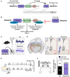
SRGAP2C as a human-specific modifier cortical connectivity and function. (A) The ancestral copy SRGAP2A, which is located on chromosome 1, is present in most mammals. Duplication of SRGAP2A in the Homo lineage resulted in the emergence of multiple copies, one of which is SRGAP2C. Humans, as the only extant Homo species, are now the only species to possess this copy. (B) SRGAP2A contains an extended F-BAR domain (F-BARx), through which it binds Homer and promotes the maturation of excitatory synapses. The SH3 domain interacts with Gephyrin to promote inhibitory synapse maturation. The Rho-GAP domain limits synaptic density through Rac1. SRGAP2C is composed of the truncated F-BAR domain of SRGAP2A, with five distinct amino acid changes specific to SRGAP2C. SRGAP2C retains the ability to dimerize with SRGAP2A and inhibits all functions of the ancestral protein. (C) Expression of SRGAP2C in mouse cortical pyramidal neurons (PNs) leads to increase synaptic density. (D) SRGAP2C-induced changes in synaptic development of cortical PNs leads to a specific increase of local and long-range cortico-cortical inputs. (E) Neuronal responses to whisker input of SRGAP2C-expressing mouse cortical PNs are more reliable and more selective to the stimulus. (F) Mice humanized for SRGAP2C expression display improved learning in a cortical-dependent whisker-based texture discrimination task. Panel F modified with permission from Schmidt et al. (2021).
Figure 5
Figure 5
Altered expression of axon guidance molecules mediate rewiring of duplicated motor pathway. Motor neurons in avian RA and mammalian cortical layer 5 express the axon guidance molecules SLIT1. ROBO1 expression in the brain stem region where vocal motor neurons are located prevent SLIT1-positive axons from innervating this area. In vocal learners, duplication of the motor pathway may have resulted in the emergence of RA and layer 5 cortical neurons with lowered SLIT1 expression, enabling these neurons to connect strongly with vocal motor neurons in the brain stem, thereby mediating the emergence of the ability of song production and speech.

References

    1. Ables J. L., Breunig J. J., Eisch A. J., Rakic P. (2011). Not(ch) just development: notch signalling in the adult brain. Nat. Rev. Neurosci. 12, 269–283. 10.1038/nrn3024 - DOI - PMC - PubMed
    1. Allman J. M., Tetreault N. A., Hakeem A. Y., Manaye K. F., Semendeferi K., Erwin J. M., et al. . (2010). The von Economo neurons in frontoinsular and anterior cingulate cortex in great apes and humans. Brain Struct. Funct. 214, 495–517. 10.1007/s00429-010-0254-0 - DOI - PubMed
    1. Almécija S., Hammond A. S., Thompson N. E., Pugh K. D., Moyà-Solà S., Alba D. M. (2021). Fossil apes and human evolution. Science 372:eabb4363. 10.1126/science.abb4363 - DOI - PubMed
    1. Araya R. (2014). Input transformation by dendritic spines of pyramidal neurons. Front. Neuroanat. 8:141. 10.3389/fnana.2014.00141 - DOI - PMC - PubMed
    1. Ardesch D. J., Scholtens L. H., Li L., Preuss T. M., Rilling J. K., van den Heuvel M. P. (2019). Evolutionary expansion of connectivity between multimodal association areas in the human brain compared with chimpanzees. Proc. Natl. Acad. Sci. U.S.A. 116, 7101–7106. 10.1073/pnas.1818512116 - DOI - PMC - PubMed

Publication types