Review
. 2022 Jan 4:12:752787.
doi: 10.3389/fphar.2021.752787. eCollection 2021.

theLiTE™: A Screening Platform to Identify Compounds that Reinforce Tight Junctions

Affiliations
Review

theLiTE™: A Screening Platform to Identify Compounds that Reinforce Tight Junctions

Teresa Lopes Gomes et al. Front Pharmacol. .

Abstract

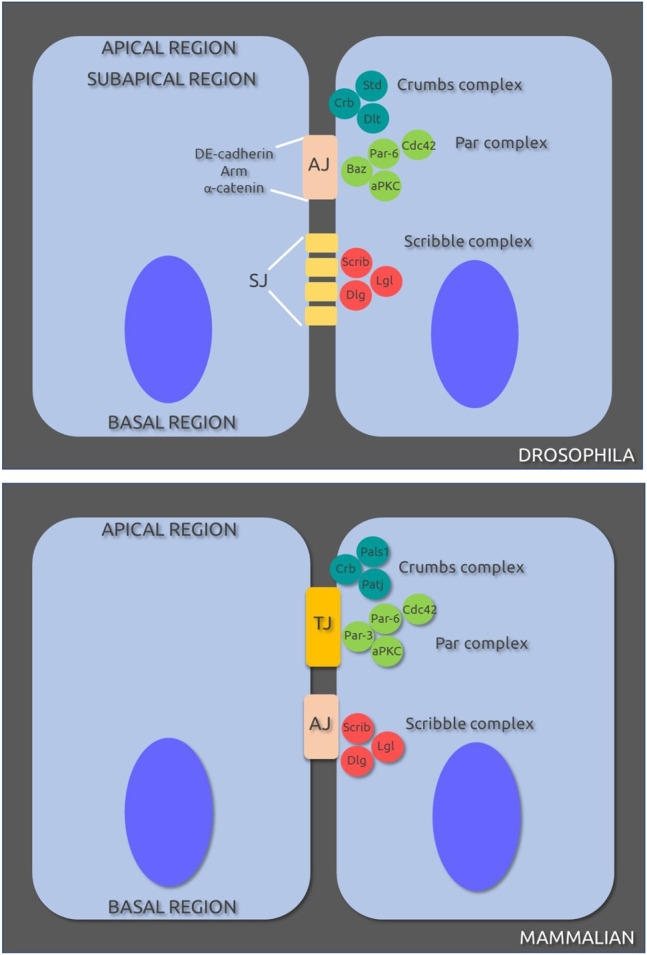
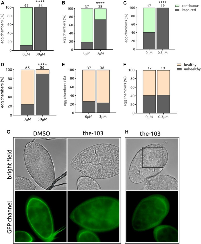
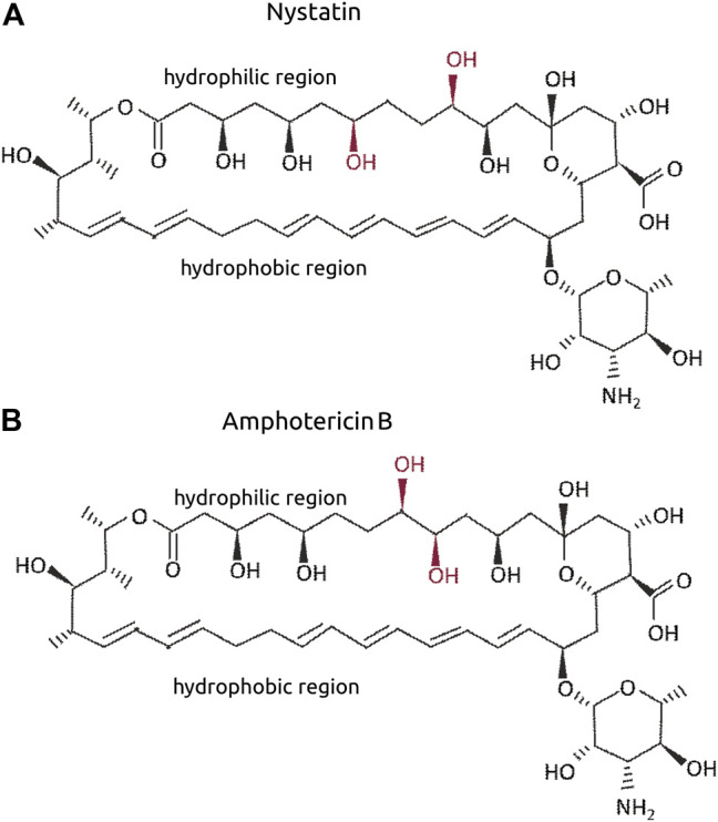
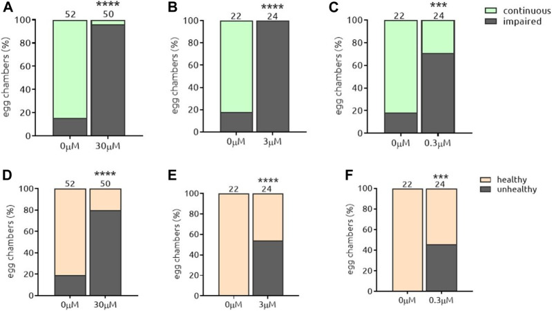
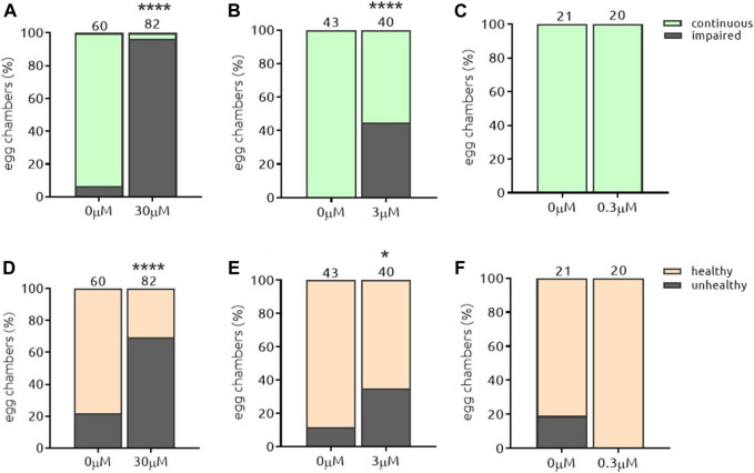
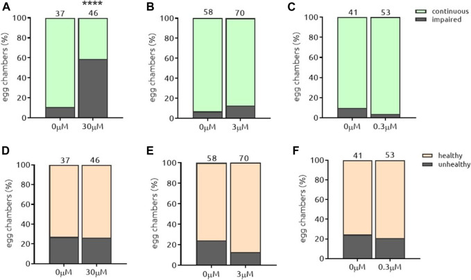
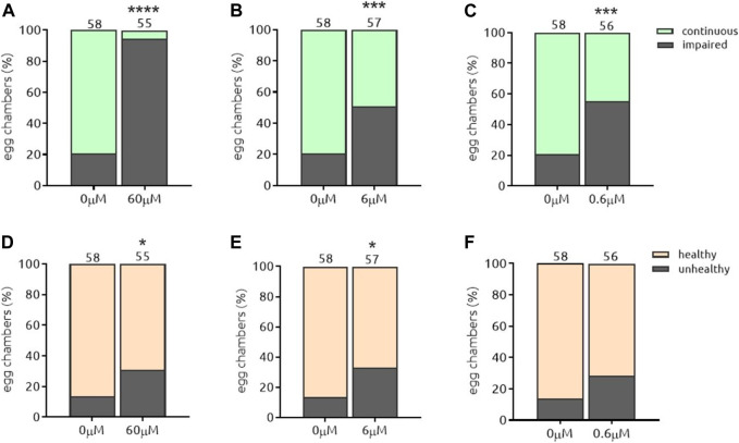
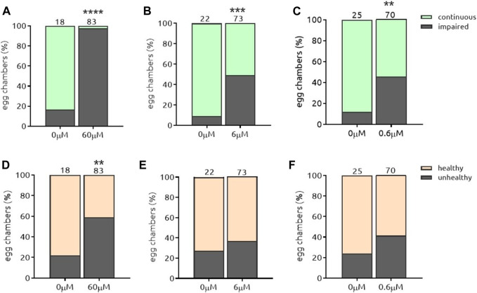
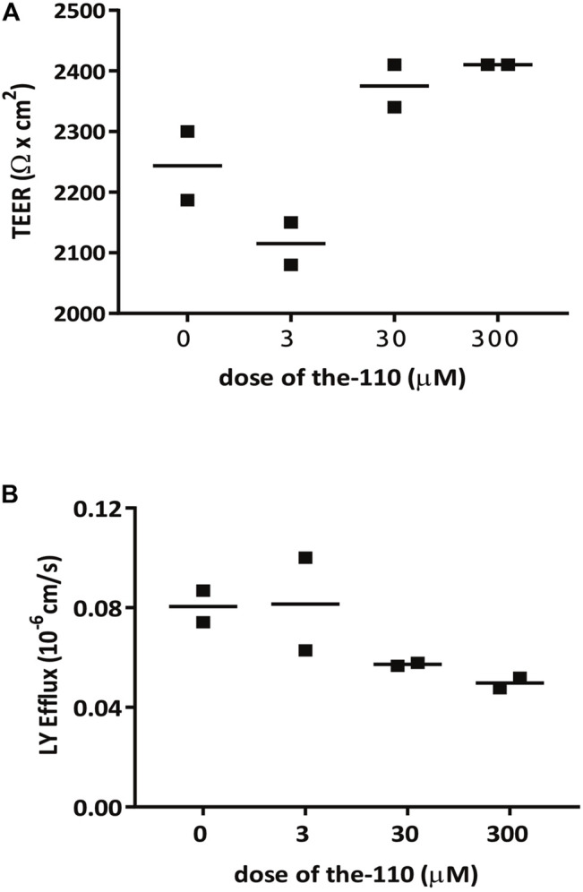
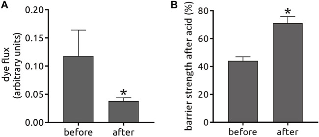
Tight junctions (TJ) are formed by transmembrane and intracellular proteins that seal the intercellular space and control selective permeability of epithelia. Integrity of the epithelial barrier is central to tissue homeostasis and barrier dysfunction has been linked to many pathological conditions. TJ support the maintenance of cell polarity through interactions with the Par complex (Cdc42-Par-6-Par-3-aPKC) in which Par-6 is an adaptor and links the proteins of the complex together. Studies have shown that Par-6 overexpression delays the assembly of TJ proteins suggesting that Par-6 negatively regulates TJ assembly. Because restoring barrier integrity is of key therapeutic and prophylactic value, we focus on finding compounds that have epithelial barrier reinforcement properties; we developed a screening platform (theLiTE™) to identify compounds that modulate Par-6 expression in follicular epithelial cells from Par-6-GFP Drosophila melanogaster egg chambers. Hits identified were then tested whether they improve epithelial barrier function, using measurements of transepithelial electrical resistance (TEER) or dye efflux to evaluate paracellular permeability. We tested 2,400 compounds, found in total 10 hits. Here we present data on six of them: the first four hits allowed us to sequentially build confidence in theLiTE™ and two compounds that were shortlisted for further development (myricetin and quercetin). We selected quercetin due to its clinical and scientific validation as a compound that regulates TJ; food supplement formulated on the basis of this discovery is currently undergoing clinical evaluation in gastroesophageal reflux disease (GERD) sufferers.

Keywords: PAR-6; epithelial cells; myricetin; polarity (cell); quercetin; tight junctions.


Conflict of interest statement

LM and RH were employed by Thelial Technologies during all or part of the work on this study. As such the funder had involvement in study design, collection, analysis, interpretation of data and the writing of this article. The decision to submit it for publication was taken independently. The remaining authors declare that the research was conducted in the absence of any commercial or financial relationships that could be construed as a potential conflict of interest.

Figures

FIGURE 1
FIGURE 1
Cell-cell junctions and protein complexes involved in polarity in Drosophila melanogaster (A) and mammalian (B) epithelial cells. AJ, adherens junctions; SJ, septate junctions; TJ, tight junctions. Original sources cited in text; for review see (Assémat et al., 2008).
FIGURE 2
FIGURE 2
Sequence of steps for automated soaking protocol. Female and male flies are put together into yeasted feeding bottles (2 female:1 male) for 2–3 days. Female flies are transferred to a CO2 pad, decapitated and processed using a blender for the isolation of egg chambers. Egg chambers are then distributed in 96-well plates, incubated with test compound for 2 hours at 25°C in the dark, without shaking. Egg chambers are washed, fixed in PFA for 15 min at RT in the dark. Fixation solution is removed and anti-fading Dabco solution added, and plate transferred to a fluorescent inverted microscope for the detection of GFP signal.
FIGURE 3
FIGURE 3
Sequence of steps for manual soaking protocol. Female and male flies are distributed into yeasted feeding bottles at a ratio of two females/1 male, for 2–3 days. Female flies were then transferred to a CO2 pad, decapitated and processed for the isolation of egg chambers. The chambers are then distributed in 0.5 ml tubes, incubated with test compound for 2 hours at 25°C in the dark without shaking. Egg chambers are washed, fixed in PFA for 15 min at RT in the dark. Fixation solution is removed and anti-fading Dabco solution added; egg chambers are then transferred to glass slide for the detection of GFP signal using a fluorescent microscope.
FIGURE 4
FIGURE 4
Par-6 ring evaluation in egg chambers. (A) Cartoon showing ovaries and egg chambers from D. melanogaster; egg chambers mature from apical towards basal. (B) Individual egg chamber showing follicular epithelium and nurse cells (nourish the oocyte). Par-6 is localized on the apical side (arrowhead) of epithelial cells (arrow) and evaluated as continuous (white arrow) (C) or impaired ring (D,E). Scale bar: 25 µm.
FIGURE 5
FIGURE 5
the-103 induces Par-6 ring impairment independent of the dose and is toxic at the highest dose. Dissected egg chambers were incubated with three different concentrations of the-103 (0.3, 3 and 30 µM) or DMSO only (0 µM) in 0.5 ml tubes for 2 h at 25°C in the dark (no shaking). Egg chambers were washed, fixed in PFA and transferred to glass slide for the detection of GFP signal using a fluorescent microscope and evaluated for Par-6 ring impairment (A–C). Epithelial and nurse cell integrity was evaluated and considered unviable by identifying the presence of a “blebbing-like” phenotype (D–E). Egg chambers containing at least one unviable cell were already considered unhealthy. (G) Example of a healthy egg chamber (bright field) incubated with the-103 showing discontinuous Par-6 ring (GFP channel). (H) Example of an unhealthy egg chamber (bright field) with faded Par-6-GFP signal. Square depicts an area of unhealthy nurse cells. ****p < 0.0001. Scale bar: 25 µm.
FIGURE 6
FIGURE 6
Molecular structure of Nystatin (A) and Amphotericin B (B). The OH in red refer to the differential positions when both are compared.
FIGURE 7
FIGURE 7
the-104a induces Par-6 ring impairment independent of the dose and is toxic at all doses. Dissected egg chambers were incubated with the-104a at 0.3, three or 30 µM or DMSO only (0 µM) in 0.5 ml tubes for 2 h at 25°C in the dark and in the absence of movement (no shaking). Egg chambers were washed, fixed and transferred to glass slide for the detection of GFP signal using a fluorescent microscope for evaluation of Par-6 ring impairment (A–C). Epithelial and nurse cell integrity was evaluated and considered unviable by identifying the presence of a “blebbing-like” phenotype (D–E). Egg chambers containing at least one unviable cell were already considered unhealthy. ***p < 0.001; ****p < 0.0001.
FIGURE 8
FIGURE 8
the-104b induces Par-6 ring impairment and toxicity in a dose-response manner. Dissected egg chambers were incubated with the-104 b for 2 h at 0.3, three or 30 µM or DMSO only (0 µM) in 0.5 ml tubes for 2 h at 25°C in the dark and in the absence of movement (no shaking). Egg chambers were washed, fixed and transferred to glass slide for the detection of GFP signal using a fluorescent microscope for evaluation of Par-6 ring impairment (A–C). Epithelial and nurse cell integrity was evaluated and considered unviable by identifying the presence of a “blebbing-like” phenotype (D–E). Egg chambers containing at least one unviable cell were already considered unhealthy. *p < 0.05; ****p < 0.0001.
FIGURE 9
FIGURE 9
the-105 induces Par-6 ring impairment only at the highest dose and is not toxic to cells. Dissected egg chambers were incubated with the-105 at three different concentrations (0.3, 3 and 30 µm) or DMSO only (0 µm) for 2 h at 25°C in 0.5 ml tubes in the dark without shaking. After incubation cells were washed, fixed with PFA and evaluated for Par-6 ring impairment (A–C). Epithelial and nurse cell integrity was evaluated and considered unviable by identifying the presence of a “blebbing-like” phenotype (D–E). Egg chambers containing at least one unviable cell were already considered unhealthy. ****p < 0.0001.
FIGURE 10
FIGURE 10
the-110 induces Par-6 ring impairment at all doses and is slightly toxic at the highest and intermediate doses. Dissected egg chambers were incubated with the-110 at concentrations of 0.6, 6 and 60 μM, or DMSO only (0 µm) in 0.5 ml tubes for 2 h at 25°C in the dark, no shaking. Cells were then washed, fixed in PFA and evaluated for Par-6 ring impairment (A–C). Epithelial and nurse cell integrity was evaluated and considered unviable by identifying the presence of a “blebbing-like” phenotype (D–E). Egg chambers containing at least one unviable cell were already considered unhealthy. *p < 0.05; ***p < 0.001; ****p < 0.0001.
FIGURE 11
FIGURE 11
the-111 induces Par-6 ring impairment at all doses and is toxic at the highest dose. Dissected egg chambers were incubated with the-111 at concentrations of 0.6, 6 and 60 μM, or DMSO only (0 µm) in 0.5 ml tubes for 2 h at 25°C in the dark, no shaking. Cells were then washed, fixed in PFA and evaluated for Par-6 ring impairment (A–C). Epithelial and nurse cell integrity was evaluated and considered unviable by identifying the presence of a “blebbing-like” phenotype (D–E). Egg chambers containing at least one unviable cell were already considered unhealthy. **p < 0.001; ***p < 0.001; ****p < 0.0001.
FIGURE 12
FIGURE 12
the-110 increases TEER and reduces paracellular permeability in Caco-2 epithelial cells. Assay was performed by Cyprotex according to their established protocol. Briefly, caco-2 cells from passage 40–60 were seeded in duplicate in a transwell insert until confluent. Cells were treated with myricetin at three different concentrations (0.3, 3 and 30 µM) or DMSO only (0 µM), in the presence of Lucifer Yellow (LY) for 2 h. Permeability was measured by transepithelial electrical resistance (TEER) and LY efflux across the barrier.
FIGURE 13
FIGURE 13
An independent open label clinical trial with quercetin was conducted in GERD patients under eligibility criteria such as age, GERD diagnosis, abnormal 24 h-pH monitoring or past responsiveness to PPI therapy (clinicaltrials.gov NCT02226484) among other criteria described in the Methods. Patients took 500 mg quercetin orally twice-a-day for 6 weeks and were monitored 1 week before and during the duration of the trial. After 6 weeks, biopsies of the esophagus were taken to determine changes in the barrier function by dye flux (A) and resistance to exposure to hydrochloric (gastric) acid (B). These results are unpublished and the use of information is disclosed under a non-exclusive license agreement between the University of North Carolina at Chapell Hill and Epinutra (Thelial BV).

References

    1. Achilleos A., Wehman A. M., Nance J. (2010). PAR-3 Mediates the Initial Clustering and Apical Localization of Junction and Polarity Proteins during C. Elegans Intestinal Epithelial Cell Polarization. Development 137 (11), 1833–1842. 10.1242/dev.047647 - DOI - PMC - PubMed
    1. Akdis C. A. (2021). Does the Epithelial Barrier Hypothesis Explain the Increase in Allergy, Autoimmunity and Other Chronic Conditions? Nat. Rev. Immunol. 21, 739–751. 10.1038/s41577-021-00538-7 - DOI - PubMed
    1. Amanzadeh E., Esmaeili A., Abadi R. E. N., Kazemipour N., Pahlevanneshan Z., Beheshti S. (2019). Quercetin Conjugated with Superparamagnetic Iron Oxide Nanoparticles Improves Learning and Memory Better Than Free Quercetin via Interacting with Proteins Involved in LTP. Sci. Rep. 9 (1), 6876. 10.1038/s41598-019-43345-w - DOI - PMC - PubMed
    1. Amasheh M., Schlichter S., Amasheh S., Mankertz J., Zeitz M., Fromm M., et al. (2008). Quercetin Enhances Epithelial Barrier Function and Increases Claudin-4 Expression in Caco-2 Cells. J. Nutr. 138 (6), 1067–1073. 10.1093/jn/138.6.1067 - DOI - PubMed
    1. Andres S., Pevny S., Ziegenhagen R., Bakhiya N., Schäfer B., Hirsch-Ernst K. I., et al. (2018). Safety Aspects of the Use of Quercetin as a Dietary Supplement. Mol. Nutr. Food Res. 62, 2018. 10.1002/mnfr.201700447 - DOI - PubMed

LinkOut - more resources