Review
. 2022 Jan 6:12:721951.
doi: 10.3389/fphys.2021.721951. eCollection 2021.

Ten Years of Pediatric Lung Ultrasound: A Narrative Review

Affiliations
Review

Ten Years of Pediatric Lung Ultrasound: A Narrative Review

Anna Maria Musolino et al. Front Physiol. .

Abstract

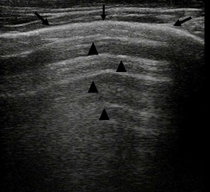
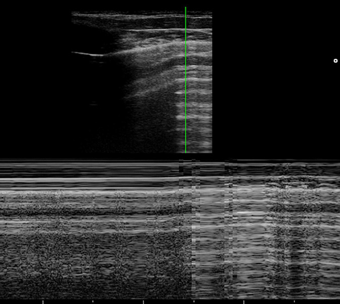
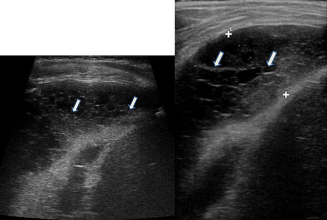
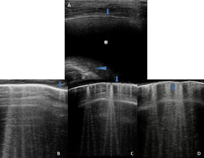
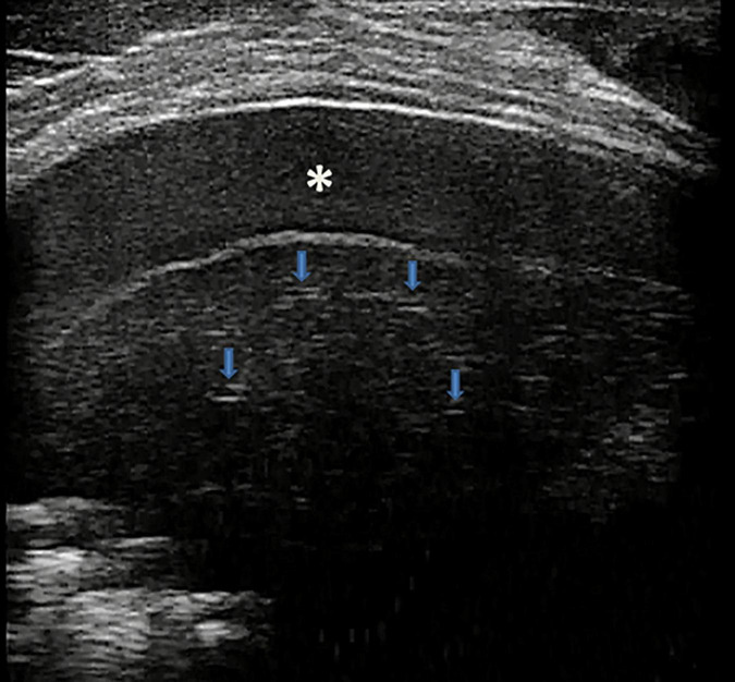
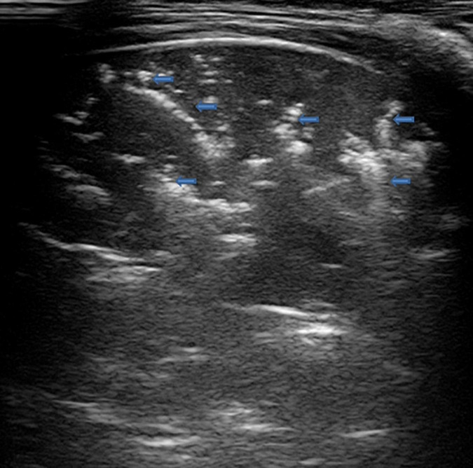
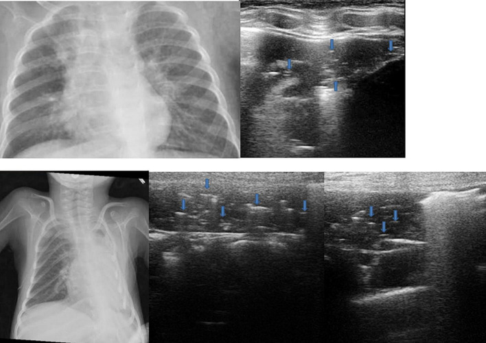
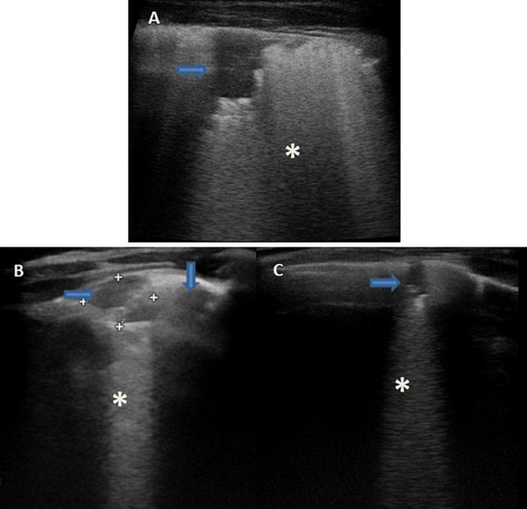
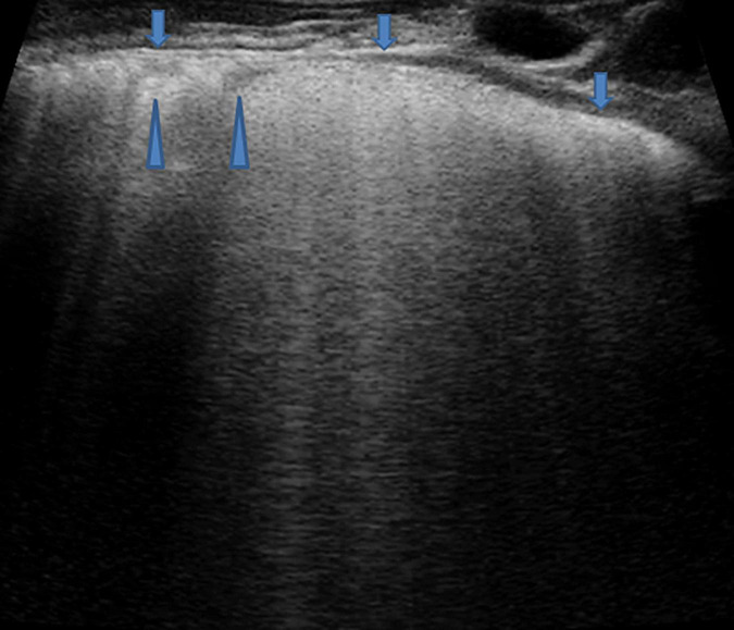
Lung diseases are the most common conditions in newborns, infants, and children and are also the primary cause of death in children younger than 5 years old. Traditionally, the lung was not thought to be a target for an ultrasound due to its inability to penetrate the gas-filled anatomical structures. With the deepening of knowledge on ultrasound in recent years, it is now known that the affected lung produces ultrasound artifacts resulting from the abnormal tissue/gas/tissue interface when ultrasound sound waves penetrate lung tissue. Over the years, the application of lung ultrasound (LUS) has changed and its main indications in the pediatric population have expanded. This review analyzed the studies on lung ultrasound in pediatrics, published from 2010 to 2020, with the aim of highlighting the usefulness of LUS in pediatrics. It also described the normal and abnormal appearances of the pediatric lung on ultrasound as well as the benefits, limitations, and possible future challenges of this modality.

Keywords: LUS; children; imaging; lung disease; lung ultrasound; pediatrics.


Conflict of interest statement

The authors declare that the research was conducted in the absence of any commercial or financial relationships that could be construed as a potential conflict of interest.

Figures

FIGURE 1
FIGURE 1
Grayscale lung ultrasound examination (transverse scan between intercostal fields; linear probe with 12 MHz frequency) shows a normal lung ultrasound pattern: hyperechoic, regular, and smooth pleural line with a thickness of less than 0.5 mm (arrows), pleural sliding present, and normally represented characterized by “to and fro” movement of lung surface synchronized with respiration (Electronic Supplementary Video 1). Below the pleural line, lung ultrasound imagines show A-lines (arrowheads): echogenic horizontal lines parallel and equidistant from each other which indicate the presence of normally aerated lung.
FIGURE 2
FIGURE 2
The grayscale lung ultrasound examination (transverse scan between the intercostal fields; linear probe with a frequency of 12 MHz) of a 15-year-old boy with left apical pneumothorax shows the Barcode sign identified in M-mode as a transition from the normal lung (linear granular pattern on the right of the figure) to the pneumothorax (linear pattern on the left of the figure). The grayscale lung ultrasound examination (transverse scan between the intercostal fields; linear probe with a frequency of 12 MHz) of a 16-year-old boy with right apical pneumothorax, shows the Lung Point sign identified in B-mode as the point where normal sliding disappears due to the presence of air in the pleural cavity.
FIGURE 3
FIGURE 3
Grayscale lung ultrasound examination (transverse scan between intercostal fields; linear probe with 12 MHz frequency) of a 5-year-old child with pneumococcal pleuropneumonia (complicated by pleural empyema), shows exudative pleural effusion, with internal echoes non-homogeneously distributed: it is fibrinous, plural-septate, and concamerated and with thickened septa (arrows).
FIGURE 4
FIGURE 4
Grayscale lung ultrasound examination (transverse scan between intercostal fields; linear probe with 12 MHz frequency) of a 10-year-old child with congestive heart disease resulting from untreated rheumatic carditis, shows in Panel (A), on the basal posterior-lateral fields on the right, an anechoic pleural effusion (asterisk) that appears like an anechoic space between the parietal (arrow) and visceral pleura (arrowhead) below which lung appears atelectasis. Panels (B,C) show the SIS pattern of the upper right and left fields, respectively; Panel (D) shows that of the right lower/basal fields: the pleural line is regular (arrows); vertical artifacts or B-lines are separated, laser-like artifacts, with gravitational course spreading from the pleural line to the bottom of the screen and they show an internal sequence of alternating horizontal bands and homogeneous distribution in the affected spaces. Ultrasound picture compatible with the septal pattern of early CPE (Soldati and Demi, 2017; Soldati et al., 2019). CPE, cardiogenic pulmonary edema; SIS, sonographic interstitial syndrome.
FIGURE 5
FIGURE 5
Grayscale lung ultrasound examination (transverse scan between intercostal fields; linear probe with 12 MHz frequency) of a 7-year-old boy with bacterial lobar pneumonia, shows hepatized subpleural consolidation with fluid bronchograms (arrows) and fibrinous inflammatory reactive pleural effusion (asterisk).
FIGURE 6
FIGURE 6
Grayscale lung ultrasound examination (transverse scan between intercostal fields; linear probe with 12 MHz frequency) of a 5-year-old boy with bacterial pneumonia lobar, shows subpleural consolidation of an inflammatory/infectious nature with numerous elements of surface dynamic arborized bronchograms (arrows) – (Electronic Supplementary Video 3) and deep fluid bronchogram.
FIGURE 7
FIGURE 7
The upper part of the figure shows chest X-ray and lung ultrasound images of a 7-year-old girl with uncontrolled asthma and an ongoing severe asthma attack, with no clinical or laboratory signs of infections. The chest X-ray shows non-specific areas of reduced transparency in the right apical and right basal sites. Grayscale lung ultrasound examination (transverse scan between intercostal fields; linear probe with 12 MHz frequency) shows – on the right paracardiac area – subpleural consolidation with hyperechoic images of air bubbles (static aerial bronchograms, arrows) that do not move inward during breathing and are parallel to each other. Their presence is associated with the identification of atelectasis, supported by the clinical and laboratory context of the patient being examined. The lower part of the figure shows chest X-ray and lung ultrasound images of a 4-year-old girl with neuromuscular pathology in nocturnal non-invasive ventilation with ongoing respiratory exacerbation without laboratory and clinical signs of infections. The chest X-ray shows a completely white left lung not well definable. Grayscale lung ultrasound examination (transverse scan between intercostal fields; linear probe with 12 MHz frequency) shows the entire left lung’s subpleural consolidations with hyperechoic images of air bubbles (static aerial bronchograms, arrows) static and parallel to each other. Their presence is associated with the identification of left pulmonary atelectasis. The ultrasound suspicion is supported by the clinical and laboratory context of the patient being examined.
FIGURE 8
FIGURE 8
Grayscale lung ultrasound examination (transverse scan between intercostal fields; linear probe with 12 MHz frequency) of children with viral lower respiratory tract infection [(A): 2-year-old boy with H1N1 Influenza pneumonia; (B,C): 8-month-old infant with Respiratory Syncytial Virus bronchiolitis]. Lung ultrasound findings show sonographic interstitial syndrome (SIS) with areas of the white lung with multiple, coalescent vertical artifacts (B-lines, asterisks) and small subpleural consolidations (hypoechoic areas, arrows) less than 1 centimeter in size associated with areas of “white lung” or confluent B-lines (asterisks).
FIGURE 9
FIGURE 9
Grayscale lung ultrasound examination (transverse scan between intercostal fields; linear probe with 12 MHz frequency) of a 4-year-old boy with viral pneumonia – due to Coronavirus (non-COVID-19), Bocavirus, and Metapneumovirus coinfection- requiring respiratory assistance with High- flow nasal oxygen at the pediatric department. It shows sonographic interstitial syndrome (SIS) which is characterized by blurred, uneven, coalescent B-lines and white lung; irregular pleural line (arrows); reduced pleural sliding; multifocal inhomogeneous involvement; subpleural microconsolidations (generating pseudo-B-lines) (arrowheads).

References

    1. Ambroggio L., Sucharew H., Rattan M. S., O’Hara S. M., Babcock D. S., Clohessy C., et al. (2016). Lung Ultrasonography: A Viable Alternative to Chest Radiography in Children with Suspected Pneumonia? J. Pediatr. 176 93.e–98.e. 10.1016/j.jpeds.2016.05.033 - DOI - PubMed
    1. Ashton-Cleary D. T. (2013). Is thoracic ultrasound a viable alternative to conventional imaging in the critical care setting? Br. J. Anaesth. 111 152–160. 10.1093/bja/aet076 - DOI - PubMed
    1. Audette L. D., Parent M. C. (2016). BET 3: Bedside lung ultrasound for the diagnosis of pneumonia in children. Emerg. Med. J. EMJ 33 589–592. 10.1136/emermed-2016-206047.3 - DOI - PubMed
    1. Avery M. E., Gatewood O. B., Brumley G. (1966). Transient tachypnea of newborn. Possible delayed resorption of fluid at birth. Am. J. Dis. Children 111 380–385. 10.1001/archpedi.1966.02090070078010 - DOI - PubMed
    1. Balk D. S., Lee C., Schafer J., Welwarth J., Hardin J., Novack V., et al. (2018). Lung ultrasound compared to chest X-ray for diagnosis of pediatric pneumonia: A meta-analysis. Pediatr. Pulmonol. 53 1130–1139. 10.1002/ppul.24020 - DOI - PubMed