

Principles of Spatial Transcriptomics Analysis: A Practical Walk-Through in Kidney Tissue
- PMID: 35069263
- PMCID: PMC8770822
- DOI: 10.3389/fphys.2021.809346
Principles of Spatial Transcriptomics Analysis: A Practical Walk-Through in Kidney Tissue
Abstract
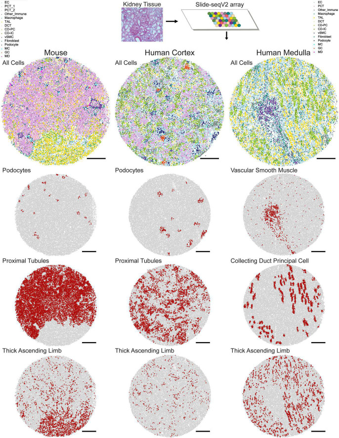
Spatial transcriptomic technologies capture genome-wide readouts across biological tissue space. Moreover, recent advances in this technology, including Slide-seqV2, have achieved spatial transcriptomic data collection at a near-single cell resolution. To-date, a repertoire of computational tools has been developed to discern cell type classes given the transcriptomic profiles of tissue coordinates. Upon applying these tools, we can explore the spatial patterns of distinct cell types and characterize how genes are spatially expressed within different cell type contexts. The kidney is one organ whose function relies upon spatially defined structures consisting of distinct cellular makeup. Thus, the application of Slide-seqV2 to kidney tissue has enabled us to elucidate spatially characteristic cellular and genetic profiles at a scale that remains largely unexplored. Here, we review spatial transcriptomic technologies, as well as computational approaches for cell type mapping and spatial cell type and transcriptomic characterizations. We take kidney tissue as an example to demonstrate how the technologies are applied, while considering the nuances of this architecturally complex tissue.
Keywords: kidney spatial transcriptomics; kidney transcriptomics; slide-seq; slide-seqV2; spatial transcriptomics.
Copyright © 2022 Noel, Wang, Greka and Marshall.
Conflict of interest statement
The authors declare that the research was conducted in the absence of any commercial or financial relationships that could be construed as a potential conflict of interest.
Figures
Similar articles
-
Highly sensitive spatial transcriptomics at near-cellular resolution with Slide-seqV2.Nat Biotechnol. 2021 Mar;39(3):313-319. doi: 10.1038/s41587-020-0739-1. Epub 2020 Dec 7. Nat Biotechnol. 2021. PMID: 33288904 Free PMC article.
-
High-resolution Slide-seqV2 spatial transcriptomics enables discovery of disease-specific cell neighborhoods and pathways.iScience. 2022 Mar 16;25(4):104097. doi: 10.1016/j.isci.2022.104097. eCollection 2022 Apr 15. iScience. 2022. PMID: 35372810 Free PMC article.
-
Identification and Localization of Cell Types in the Mouse Olfactory Bulb Using Slide-SeqV2.Methods Mol Biol. 2023;2710:171-183. doi: 10.1007/978-1-0716-3425-7_13. Methods Mol Biol. 2023. PMID: 37688732 Free PMC article.
-
Computational solutions for spatial transcriptomics.Comput Struct Biotechnol J. 2022 Sep 1;20:4870-4884. doi: 10.1016/j.csbj.2022.08.043. eCollection 2022. Comput Struct Biotechnol J. 2022. PMID: 36147664 Free PMC article. Review.
-
Computational exploration of cellular communication in skin from emerging single-cell and spatial transcriptomic data.Biochem Soc Trans. 2022 Feb 28;50(1):297-308. doi: 10.1042/BST20210863. Biochem Soc Trans. 2022. PMID: 35191953 Free PMC article. Review.
Cited by
-
The dawn of spatial omics.Science. 2023 Aug 4;381(6657):eabq4964. doi: 10.1126/science.abq4964. Epub 2023 Aug 4. Science. 2023. PMID: 37535749 Free PMC article. Review.
-
Reshaping transplantation with AI, emerging technologies and xenotransplantation.Nat Med. 2025 Jul;31(7):2161-2173. doi: 10.1038/s41591-025-03801-9. Epub 2025 Jul 14. Nat Med. 2025. PMID: 40659768 Review.
-
Artifacts in spatial transcriptomics data: their detection, importance, prevalence, and prevention.Brief Bioinform. 2025 Jul 2;26(4):bbaf306. doi: 10.1093/bib/bbaf306. Brief Bioinform. 2025. PMID: 40748322 Free PMC article.
-
Novel insights into kidney disease: the scRNA-seq and spatial transcriptomics approaches: a literature review.BMC Nephrol. 2025 Apr 8;26(1):181. doi: 10.1186/s12882-025-04103-5. BMC Nephrol. 2025. PMID: 40200175 Free PMC article. Review.
-
Deep learning in spatial transcriptomics: Learning from the next next-generation sequencing.Biophys Rev (Melville). 2023 Feb 7;4(1):011306. doi: 10.1063/5.0091135. eCollection 2023 Mar. Biophys Rev (Melville). 2023. PMID: 38505815 Free PMC article. Review.
References
-
- 10x Genomics (2007). Spatial Gene Expression – 10x Genomics. Available online at: https://www.10xgenomics.com/products/spatial-gene-expression (accessed October 31, 2021).
-
- Bellock K., Godber N., Kahn P. (2021). bellockk/alphashape: v1.3.1 Release. Zenodo. 10.5281/ZENODO.4697576 - DOI
Publication types
LinkOut - more resources
Full Text Sources