Randomized Controlled Trial
. 2022 Jan 5:12:798207.
doi: 10.3389/fimmu.2021.798207. eCollection 2021.

Induction of Functional Specific Antibodies, IgG-Secreting Plasmablasts and Memory B Cells Following BCG Vaccination

Affiliations
Randomized Controlled Trial

Induction of Functional Specific Antibodies, IgG-Secreting Plasmablasts and Memory B Cells Following BCG Vaccination

Julia Bitencourt et al. Front Immunol. .

Abstract

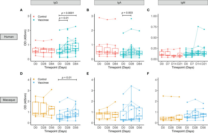
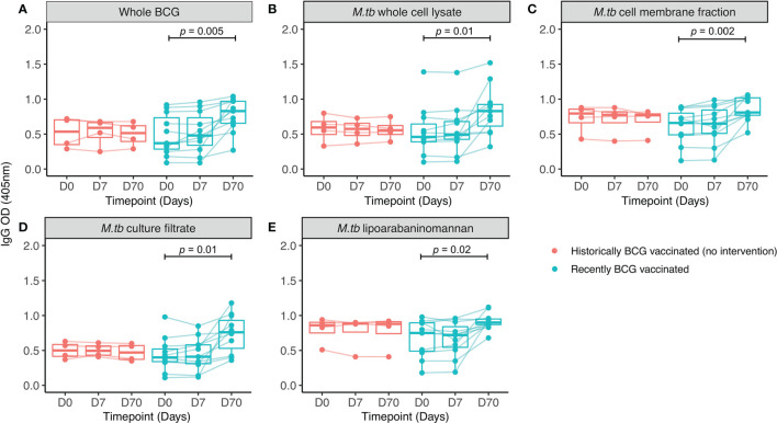
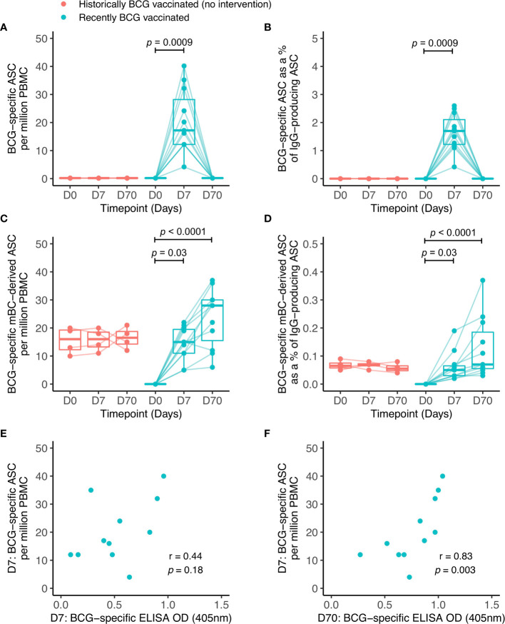
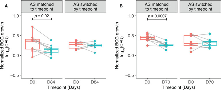
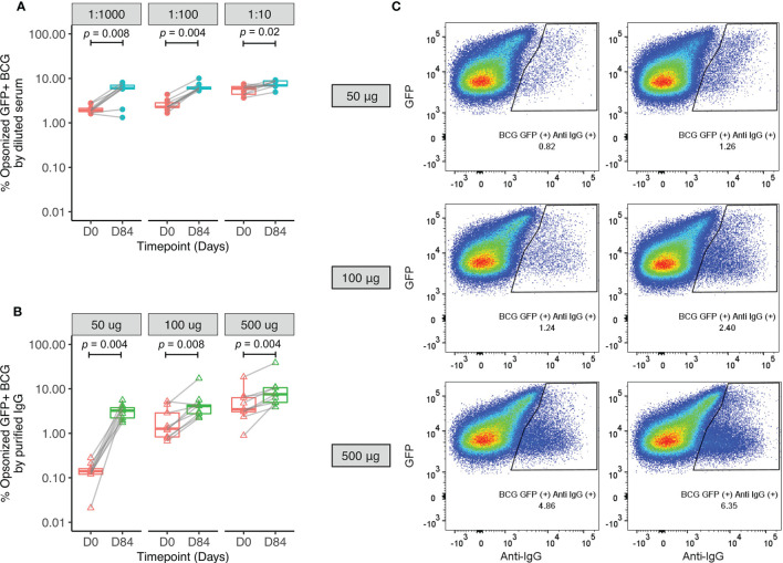
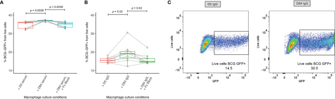
Tuberculosis (TB) is a major global health problem and the only currently-licensed vaccine, BCG, is inadequate. Many TB vaccine candidates are designed to be given as a boost to BCG; an understanding of the BCG-induced immune response is therefore critical, and the opportunity to relate this to circumstances where BCG does confer protection may direct the design of more efficacious vaccines. While the T cell response to BCG vaccination has been well-characterized, there is a paucity of literature on the humoral response. We demonstrate BCG vaccine-mediated induction of specific antibodies in different human populations and macaque species which represent important preclinical models for TB vaccine development. We observe a strong correlation between antibody titers in serum versus plasma with modestly higher titers in serum. We also report for the first time the rapid and transient induction of antibody-secreting plasmablasts following BCG vaccination, together with a robust and durable memory B cell response in humans. Finally, we demonstrate a functional role for BCG vaccine-induced specific antibodies in opsonizing mycobacteria and enhancing macrophage phagocytosis in vitro, which may contribute to the BCG vaccine-mediated control of mycobacterial growth observed. Taken together, our findings indicate that the humoral immune response in the context of BCG vaccination merits further attention to determine whether TB vaccine candidates could benefit from the induction of humoral as well as cellular immunity.

Keywords: B cells; BCG; TB; antibodies; humoral immunity; tuberculosis; vaccine.


Conflict of interest statement

The authors declare that the research was conducted in the absence of any commercial or financial relationships that could be construed as a potential conflict of interest.

Figures

Figure 1
Figure 1
PPD-specific antibody responses to BCG vaccination in healthy UK adults and rhesus macaques. Serum was collected from volunteers enrolled into human Study 1 who were either unvaccinated controls (red) or received BCG vaccination (cyan) (A–C), and from rhesus macaques enrolled into macaque Study 1 which were either unvaccinated controls (yellow) or received BCG vaccination (blue) (D–F). PPD-specific IgG (A, D), IgA (B, E) and IgM (C, F) responses were measured over time. Points represent the mean of triplicate values, boxes indicate the median value with the interquartile range (IQR) and the upper whisker extends to the largest value no further than 1.5 * IQR from the hinge, the lower whisker extends from the hinge to the smallest value at most 1.5 * IQR from the hinge. Outliers are plotted individually. A mixed effects analysis with Dunnett’s correction for multiple comparisons was conducted on logged values (A–C) or a Friedman test with Dunn’s correction for multiple comparisons (D–F) was performed to compare the BCG vaccine-induced response between post-vaccination and baseline time-points.
Figure 2
Figure 2
IgG responses to different mycobacterial fractions in serum collected from Nepalese military recruits. Serum samples were taken from Nepalese adults enrolled into human Study 2 (red = historically BCG-vaccinated individuals who received no intervention, cyan = BCG-naïve individuals who received BCG vaccination). IgG responses to whole BCG (A), M.tb whole cell lysate (B), M.tb cell membrane fraction (C), M.tb culture filtrate (D) or M.tb lipoarabaninomannan (E) were measured. Points represent the mean of triplicate values, boxes indicate the median value with the interquartile range (IQR) and the upper whisker extends to the largest value no further than 1.5 * IQR from the hinge, the lower whisker extends from the hinge to the smallest value at most 1.5 * IQR from the hinge. Outliers are plotted individually. A repeated measures one-way ANOVA with Dunnett’s multiple comparisons test was used to compare between time-points in the BCG vaccinated group.
Figure 3
Figure 3
BCG-specific IgG-secreting plasmablast (ASC) and memory B cell-derived ASC responses following BCG vaccination. ASC and mBC-derived ASC responses were determined at baseline, 7 days and 70 days using cells collected from Nepalese military recruits enrolled into human Study 2 (red = historically BCG-vaccinated individuals who received no intervention, cyan = BCG-naïve individuals who received BCG vaccination). ASC responses are presented as the number of BCG-specific ASCs per million PBMC (A) and as the proportion (%) of total IgG-secreting ASC (B). mBC responses are shown as the number of BCG-specific mBC-derived ASC per million cultured PBMC (C) and as the proportion (%) of mBC-derived ASC of total IgG-secreting ASC (D). Points represent the mean of triplicate values, boxes indicate the median value with the interquartile range (IQR) and the upper whisker extends to the largest value no further than 1.5 * IQR from the hinge, the lower whisker extends from the hinge to the smallest value at most 1.5 * IQR from the hinge. Outliers are plotted individually. A Friedman test with Dunn’s multiple comparisons test was used (A–D). A Spearman rank correlation was performed between ASC responses and BCG-specific IgG responses measured at 7 days (E) and 70 days (F) after BCG vaccination.
Figure 4
Figure 4
Serum factors contribute to BCG-induced control of mycobacterial growth in vitro. Samples were used from n=9 healthy UK adults enrolled into human Study 1 (A) and n=9 healthy Nepalese military recruits enrolled into human Study 2 (B), all of whom received ID BCG vaccination. The direct MGIA was performed using PBMC collected at baseline and 84 days (A) or 70 days (B) post-BCG vaccination, co-cultured with BCG and either autologous serum matched to time-point, or autologous serum switched by time-point ie. post-vaccination serum cultured with baseline PBMC and baseline serum cultured with post-vaccination PBMC. Normalized BCG growth = (log10 CFU of sample - log10 CFU of growth control). Points represent the mean of duplicate values, boxes indicate the median value with the interquartile range (IQR) and the upper whisker extends to the largest value no further than 1.5 * IQR from the hinge, the lower whisker extends from the hinge to the smallest value at most 1.5 * IQR from the hinge. Outliers are plotted individually. A paired t-test was used to compare between time-points.
Figure 5
Figure 5
Enhanced opsonization of BCG-GFP with sera or purified IgG following BCG vaccination. Samples were used from n=9 healthy UK adults enrolled into human Study 1. BCG-GFP was incubated with either serum at different dilutions (A) or purified IgG at different concentrations (B) collected at baseline and 84 days post-BCG vaccination, and the percentage of BCG-GFP that was opsonized was determined by flow cytometry. Points represent the mean of duplicate values, boxes indicate the median value with the interquartile range (IQR) and the upper whisker extends to the largest value no further than 1.5 * IQR from the hinge, the lower whisker extends from the hinge to the smallest value at most 1.5 * IQR from the hinge. Outliers are plotted individually. A Wilcoxon matched-pairs test was used to compare between time-points. Flow plots are shown from a single replicate of one subject at baseline and 84 days post-BCG vaccination using different concentrations of purified IgG (C).
Figure 6
Figure 6
Enhanced macrophage phagocytosis of BCG-GFP with sera or purified IgG following BCG vaccination. Samples were used from n=9 healthy UK adults enrolled into human Study 1. Antibody-dependent cellular phagocytosis was measured using human THP-1 derived macrophages and serum (A) or purified IgG (B) from baseline and 84 days post-BCG vaccination and the percentage of BCG-GFP that was phagocytosed was determined by flow cytometry. Points represent the mean of duplicate values, boxes indicate the median value with the interquartile range (IQR) and the upper whisker extends to the largest value no further than 1.5 * IQR from the hinge, the lower whisker extends from the hinge to the smallest value at most 1.5 * IQR from the hinge. Outliers are plotted individually. A Wilcoxon matched-pairs test was used to compare between time-points. Flow plots are shown from a single replicate of one subject at baseline and 84 days post-BCG vaccination using purified IgG (C).

References

    1. WHO . World Health Organisation Global Tuberculosis Report 2020. Geneva:World Health Organization; (2020).
    1. Orme IM. Characteristics and Specificity of Acquired Immunologic Memory to Mycobacterium Tuberculosis Infection. J Immunol (1988) 140(10):3589–93. - PubMed
    1. Jasenosky LD, Scriba TJ, Hanekom WA, Goldfeld AE. T Cells and Adaptive Immunity to Mycobacterium Tuberculosis in Humans. Immunol Rev (2015) 264(1):74–87. doi: 10.1111/imr.12274 - DOI - PubMed
    1. Caruso AM, Serbina N, Klein E, Triebold K, Bloom BR, Flynn JL. Mice Deficient in CD4 T Cells Have Only Transiently Diminished Levels of IFN-Gamma, Yet Succumb to Tuberculosis. J Immunol (1999) 162(9):5407–16. - PubMed
    1. Flynn JL, Chan J, Triebold KJ, Dalton DK, Stewart TA, Bloom BR. An Essential Role for Interferon Gamma in Resistance to Mycobacterium Tuberculosis Infection. J Exp Med (1993) 178(6):2249–54. doi: 10.1084/jem.178.6.2249 - DOI - PMC - PubMed

Publication types

MeSH terms

LinkOut - more resources