. 2022 Jan 7:12:797747.
doi: 10.3389/fgene.2021.797747. eCollection 2021.

Single-Cell RNA Profiling of Human Skin Reveals Age-Related Loss of Dermal Sheath Cells and Their Contribution to a Juvenile Phenotype

Affiliations

Single-Cell RNA Profiling of Human Skin Reveals Age-Related Loss of Dermal Sheath Cells and Their Contribution to a Juvenile Phenotype

Juliane M D Ahlers et al. Front Genet. .

Abstract

The dermal sheath (DS) is a population of mesenchyme-derived skin cells with emerging importance for skin homeostasis. The DS includes hair follicle dermal stem cells, which exhibit self-renewal and serve as bipotent progenitors of dermal papilla (DP) cells and DS cells. Upon aging, stem cells exhibit deficiencies in self-renewal and their number is reduced. While the DS of mice has been examined in considerable detail, our knowledge of the human DS, the pathways contributing to its self-renewal and differentiation capacity and potential paracrine effects important for tissue regeneration and aging is very limited. Using single-cell RNA sequencing of human skin biopsies from donors of different ages we have now analyzed the transcriptome of 72,048 cells, including 50,149 fibroblasts. Our results show that DS cells that exhibit stem cell characteristics were lost upon aging. We further show that HES1, COL11A1, MYL4 and CTNNB1 regulate DS stem cell characteristics. Finally, the DS secreted protein Activin A showed paracrine effects on keratinocytes and dermal fibroblasts, promoting proliferation, epidermal thickness and pro-collagen production. Our work provides a detailed description of human DS identity on the single-cell level, its loss upon aging, its stem cell characteristics and its contribution to a juvenile skin phenotype.

Keywords: Activin A; aging; dermal sheath cells; fibroblasts; regeneration; single-cell RNA sequencing; skin; stem cells.


Conflict of interest statement

Authors JMDA, CF, NH, HW, MW, EG and AS are employed by the Beiersdorf AG. The remaining authors declare that the research was conducted in the absence of any commercial or financial relationships that could be construed as a potential conflict of interest.

Figures
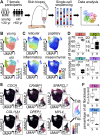
FIGURE 1
FIGURE 1
The cellular composition of dermal fibroblasts changes upon aging. (A) Experimental design. (B) Uniform Manifold Approximation and Projection (UMAP) plots of fibroblasts of young and old participants. (C) Expression of reticular (WISP2, SLPI, CTHRC1, MFAP5, TSPAN8), papillary (APCDD1, ID1, WIF1, COL18A1, PTGDS), pro-inflammatory (CCL19, APOE, CXCL2, CXCL3, EFEMP1) and mesenchymal (ASPN, POSTN, GPC3, TNN, SFRP1) markers (Solé-Boldo et al., 2020) among fibroblasts in UMAP plots. (D) Box plot of percentages of cells in fibroblast populations young vs. old, **** p<0.0001 (Fisher’s exact test). (E) Marker expression in UMAP plots, subclustering of population 4 into 4a and 4b. (F) Box plot of percentages of cells in fibroblast populations 4a and 4b young, vs. old, **** p<0.0001 (Fisher's exact test). F1-F4b Fibroblast populations 1–4b, yr years, y young, o old.
FIGURE 2
FIGURE 2
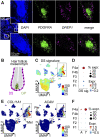
Subpopulation 4a represents the dermal sheath (DS). (A) RNA-FISH co-staining of subpopulation 4a marker DPEP1 with PDGFRA, n = 5. (B) Mesoderm-derived cell types of the hair follicle: DS cells, dermal papilla (DP) cells. Average expression of top 10 DS signature genes (CCDC80, COL11A1, TNMD, LRRC15, COL12A1, FGL2, CD200, FAM101B, SPARC, IGFBP7) (Shin et al., 2020) (C) among all cell types in Uniform Manifold Approximation and Projection (UMAP) plot and (D) among fibroblast populations in dot plot. (E) Expression of DS-specific genes COL11A1 and ACAN among all cell types in UMAP plots. (F) Dot plot of the expression of DS marker genes ACTA2 (A) and TAGLN (T) among fibroblast populations. % expr. Percent expressed, expr. average expression, Mel melanocytes, Per pericytes.
FIGURE 3
FIGURE 3
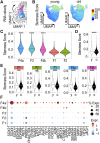
Validation of Dermal sheath (DS) population and its loss upon aging in independent scRNA-seq datasets. Uniform Manifold Approximation and Projection (UMAP) plots of fibroblast populations from (A) Vorstandlechner et al., 2020, (B) Rojahn et al., 2020, (C) Tabib et al., 2020 and (D) Solé-Boldo et al., 2020. Violin plots of the average expression of the ten most upregulated genes of DS population (COL11A1, DPEP1, POSTN, TAGLN, MEF2C, MYL4, TNMD, WFDC1, GPC3, PPP1R14A) in these datasets. (E) UMAP plot of fibroblasts of other datasets integrated together with our dataset. Percentage of DS population young vs. old, n = 8 (young), n = 9 (old). *p < 0.05 (two-tailed unpaired t-test), mean +/- SEM. FB Fibroblasts, SR secretory-reticular, PI pro-inflammatory, SP secretory-papillary, M mesenchymal.
FIGURE 4
FIGURE 4
The dermal sheath population likely contains progenitor cells that are lost upon aging. (A) RNA velocity analysis of mesenchymal population 4, computed arrows indicating the extrapolated future states of the cells, bold hand-drawn arrows indicating 2 cell fates. (B) Stemness score of each cell in Uniform Manifold Approximation and Projection (UMAP) plots young vs. old. Violin plots of stemness score of (C) young populations, (D) young vs. old fibroblasts and (E) young vs. old populations. ***p < 0.001, ****p < 0.0001 (unpaired Wilcoxon rank sum tests). (F) Dot plot of the expression of genes accounting for “stemness” gene ontology terms by fibroblast populations. F1-4b Fibroblast populations 1–4b, y young, o old. % expr. Percent expressed, expr. average expression.
FIGURE 5
FIGURE 5
Characteristic genes of the dermal sheath regulate stem cell characteristics. (A) Adipogenic differentiation potential, (B) chondrogenic differentiation potential and (C) proliferation, 72 h after knockdown of DS genes. n = 5, *p < 0.05, **p < 0.01, ***p < 0.001, ****p < 0.0001 (RM one-way ANOVA with Dunnett multiple testing correction).
FIGURE 6
FIGURE 6
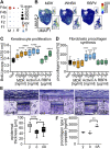
Secreted proteins of the dermal sheath mediate paracrine effects on fibroblasts and keratinocytes and contribute to a juvenile skin phenotype. Expression of INHBA (I), MDK (M) and RBP4 (R) among the fibroblast populations (A) in dot plot and (B) in Uniform Manifold Approximation and Projection plots. Box plots of (C) proliferation of old keratinocytes and (D) procollagen type I c-peptide synthesis of old fibroblasts after treatment with secreted proteins MDK, Activin A and RBP4 for 72 h, n = 5, *p < 0.05, **p < 0.01, ***p < 0.001, ****p < 0.0001 (RM one-way ANOVA with Dunnett multiple testing correction). (E) Epidermal thickness of 3D skin equivalents stained with Diff-Quick (microscopy images) and (F) procollagen synthesis. Comparison of young (y) vs. old (o) and old vs. old Activin A treated (100 ng/ml for 6 weeks, old A/oA) (box plots), n = 3, 5 slices were used for each n to determine epidermal thickness, *p < 0.05, **p < 0.01, ***p < 0.001, ****p < 0.0001 (two-tailed un/paired t-test).

References

    1. Antsiferova M., Klatte J. E., Bodó E., Paus R., Jorcano J. L., Matzuk M. M., et al. (2009). Keratinocyte-derived Follistatin Regulates Epidermal Homeostasis and Wound Repair. Lab. Invest. 89, 131–141. 10.1038/labinvest.2008.120 - DOI - PMC - PubMed
    1. Ayala-Cuellar A. P., Kang J.-H., Jeung E.-B., Choi K.-C. (2019). Roles of Mesenchymal Stem Cells in Tissue Regeneration and Immunomodulation. Biomolecules Ther. 27, 25–33. 10.4062/biomolther.2017.260 - DOI - PMC - PubMed
    1. Boehnke K., Mirancea N., Pavesio A., Fusenig N. E., Boukamp P., Stark H.-J. (2007). Effects of Fibroblasts and Microenvironment on Epidermal Regeneration and Tissue Function in Long-Term Skin Equivalents. Eur. J. Cel. Biol. 86, 731–746. 10.1016/j.ejcb.2006.12.005 - DOI - PubMed
    1. Collins C. A., Kretzschmar K., Watt F. M. (2011). Reprogramming Adult Dermis to a Neonatal State through Epidermal Activation of β-catenin. Development 138, 5189–5199. 10.1242/dev.064592 - DOI - PMC - PubMed
    1. Dominici M., Le Blanc K., Mueller I., Slaper-Cortenbach I., Marini F. C., Krause D. S., et al. (2006). Minimal Criteria for Defining Multipotent Mesenchymal Stromal Cells. The International Society for Cellular Therapy Position Statement. Cytotherapy 8, 315–317. 10.1080/14653240600855905 - DOI - PubMed