

Schwann Cells Accelerate Osteogenesis via the Mif/CD74/FOXO1 Signaling Pathway In Vitro
- PMID: 35069747
- PMCID: PMC8776480
- DOI: 10.1155/2022/4363632
Schwann Cells Accelerate Osteogenesis via the Mif/CD74/FOXO1 Signaling Pathway In Vitro
Abstract
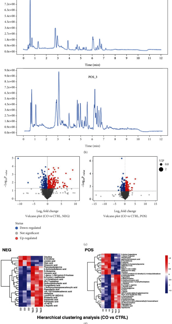
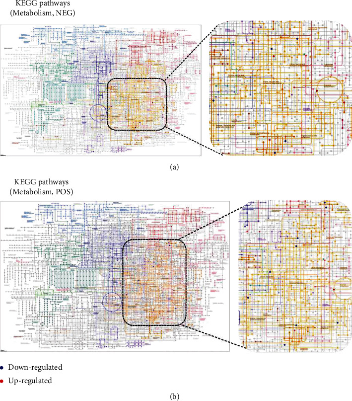
Schwann cells have been found to promote osteogenesis by an unclear molecular mechanism. To better understand how Schwann cells accelerate osteogenesis, RNA-Seq and LC-MS/MS were utilized to explore the transcriptomic and metabolic response of MC3T3-E1 to Schwann cells. Osteogenic differentiation was determined by ALP staining. Lentiviruses were constructed to alter the expression of Mif (macrophage migration inhibitory factor) in Schwann cells. Western blot (WB) analysis was employed to detect the protein expression. The results of this study show that Mif is essential for Schwann cells to promote osteogenesis, and its downstream CD74/FOXO1 is also involved in the promotion of Schwann cells on osteogenesis. Further, Schwann cells regulate amino acid metabolism and lipid metabolism in preosteoblasts. These findings unveil the mechanism for Schwann cells to promote osteogenesis where Mif is a key factor.
Copyright © 2022 Jun-Qin Li et al.
Conflict of interest statement
The authors declare no competing interests.
Figures


Similar articles
-
MIF/CD74 axis participates in inflammatory activation of Schwann cells following sciatic nerve injury.J Mol Histol. 2019 Aug;50(4):355-367. doi: 10.1007/s10735-019-09832-0. Epub 2019 Jun 13. J Mol Histol. 2019. PMID: 31197516
-
Deletion of CD74, a putative MIF receptor, in mice enhances osteoclastogenesis and decreases bone mass.J Bone Miner Res. 2013 Apr;28(4):948-59. doi: 10.1002/jbmr.1787. J Bone Miner Res. 2013. PMID: 23044992 Free PMC article.
-
Differential circRNA expression profiles during the BMP2-induced osteogenic differentiation of MC3T3-E1 cells.Biomed Pharmacother. 2017 Jun;90:492-499. doi: 10.1016/j.biopha.2017.03.051. Epub 2017 Apr 7. Biomed Pharmacother. 2017. PMID: 28395271
-
Macrophage migration inhibitory factor and CD74 regulate macrophage chemotactic responses via MAPK and Rho GTPase.J Immunol. 2011 Apr 15;186(8):4915-24. doi: 10.4049/jimmunol.1003713. Epub 2011 Mar 16. J Immunol. 2011. PMID: 21411731 Free PMC article.
-
CD74 in Kidney Disease.Front Immunol. 2015 Sep 23;6:483. doi: 10.3389/fimmu.2015.00483. eCollection 2015. Front Immunol. 2015. PMID: 26441987 Free PMC article. Review.
Cited by
-
Small Intestinal Submucosa Biomimetic Periosteum Promotes Bone Regeneration.Membranes (Basel). 2022 Jul 20;12(7):719. doi: 10.3390/membranes12070719. Membranes (Basel). 2022. PMID: 35877922 Free PMC article.
-
Novel Cinnamaldehyde Derivatives Inhibit Peripheral Nerve Degeneration by Targeting Schwann Cells.Antioxidants (Basel). 2022 Sep 20;11(10):1846. doi: 10.3390/antiox11101846. Antioxidants (Basel). 2022. PMID: 36290569 Free PMC article.
References
-
- Wu Z., Pu P., Su Z., Zhang X., Nie L., Chang Y. Schwann cell-derived exosomes promote bone regeneration and repair by enhancing the biological activity of porous Ti6Al4V scaffolds. Biochemical and Biophysical Research Communications . 2020;531(4):559–565. doi: 10.1016/j.bbrc.2020.07.094. - DOI - PubMed
-
- Xie M., Kamenev D., Kaucka M., et al. Schwann cell precursors contribute to skeletal formation during embryonic development in mice and zebrafish. Proceedings of the National Academy of Sciences of the United States of America . 2019;116(30):15068–15073. doi: 10.1073/pnas.1900038116. - DOI - PMC - PubMed
-
- Zheng L., Gao J., Jin K., et al. Macrophage migration inhibitory factor (MIF) inhibitor 4-IPP suppresses osteoclast formation and promotes osteoblast differentiation through the inhibition of the NF-κB signaling pathway. The FASEB Journal . 2019;33(6):7667–7683. doi: 10.1096/fj.201802364RR. - DOI - PubMed
LinkOut - more resources
Full Text Sources
Research Materials
Miscellaneous
