

A giant basilar artery perforator aneurysm
- PMID: 35069959
- PMCID: PMC8762371
- DOI: 10.1016/j.radcr.2021.12.034
A giant basilar artery perforator aneurysm
Abstract
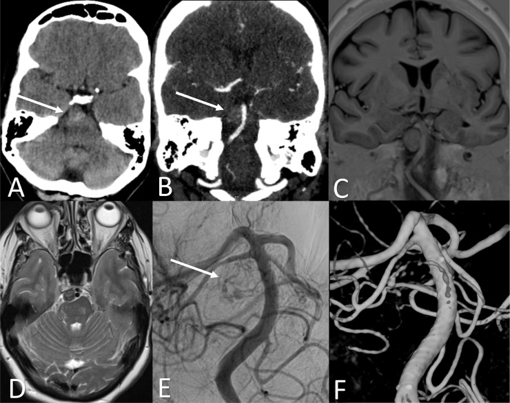
Basilar artery perforator aneurysms (BAPA's) are a rare entity. Their natural history and treatment are unclear. We describe the largest BAPA reported thus far in literature in a 64-year-old Caucasian woman. This patient did not present with subarachnoid hemorrhage, but with left hemiparesis due to pontine ischemia. The aneurysm was initially misdiagnosed as a tumoral mass in a referring center. Angiography confirmed the presence of a BAPA and a flow diverter was successfully placed. This case shows us that a BAPA can mimic a tumoral mass and can cause ischemia due to mass effect without having ruptured. Both conservative and flow diverter placement seems viable treatment options. Individual patient characteristics and preferences should be considered in decision-making for treatment.
Keywords: Intracranial Aneurysm; Stents; Subarachnoid Hemorrhage.
© 2021 The Authors. Published by Elsevier Inc. on behalf of University of Washington.
Conflict of interest statement
The manuscript has not been submitted elsewhere nor published elsewhere in whole or in part. None of the authors has potential conflicts of interest related to this manuscript.
Figures
References
-
- Chau Y, Sachet M, Sedat J. Should we treat aneurysms in perforator arteries from the basilar trunk? Review of 49 cases published in the literature and presentation of three personal cases. Interv Neuroradiol. 2018;24(1):22–28. doi: 10.1177/1591019917734531. [published Online First: 2017/10/13] - DOI - PMC - PubMed
Publication types
LinkOut - more resources
Full Text Sources
Medical
