. 2022 Jan 11:2022:4036274.
doi: 10.1155/2022/4036274. eCollection 2022.

Oxidative Stress Aggravates Apoptosis of Nucleus Pulposus Cells through m6A Modification of MAT2A Pre-mRNA by METTL16

Affiliations

Oxidative Stress Aggravates Apoptosis of Nucleus Pulposus Cells through m6A Modification of MAT2A Pre-mRNA by METTL16

Peng-Bo Chen et al. Oxid Med Cell Longev. .

Abstract

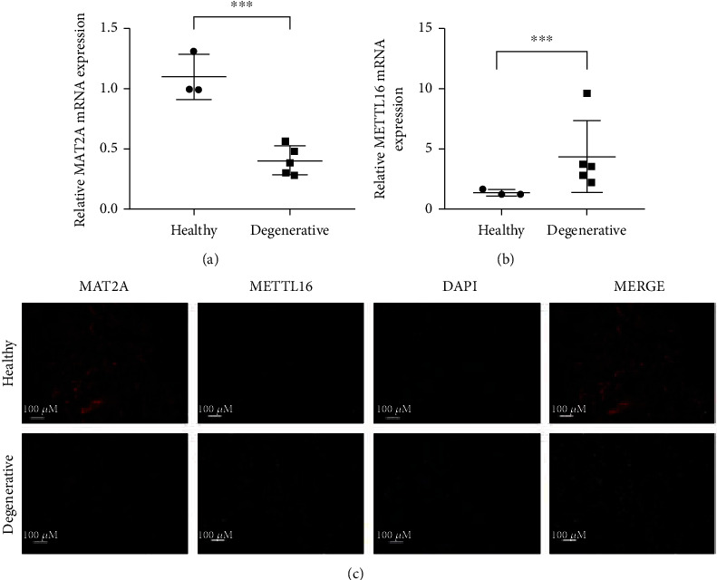
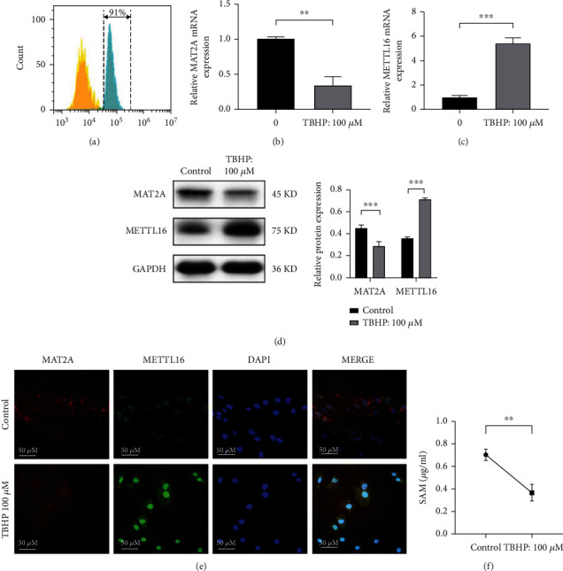
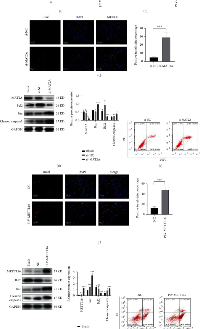
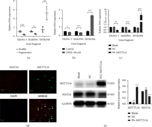
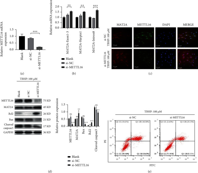
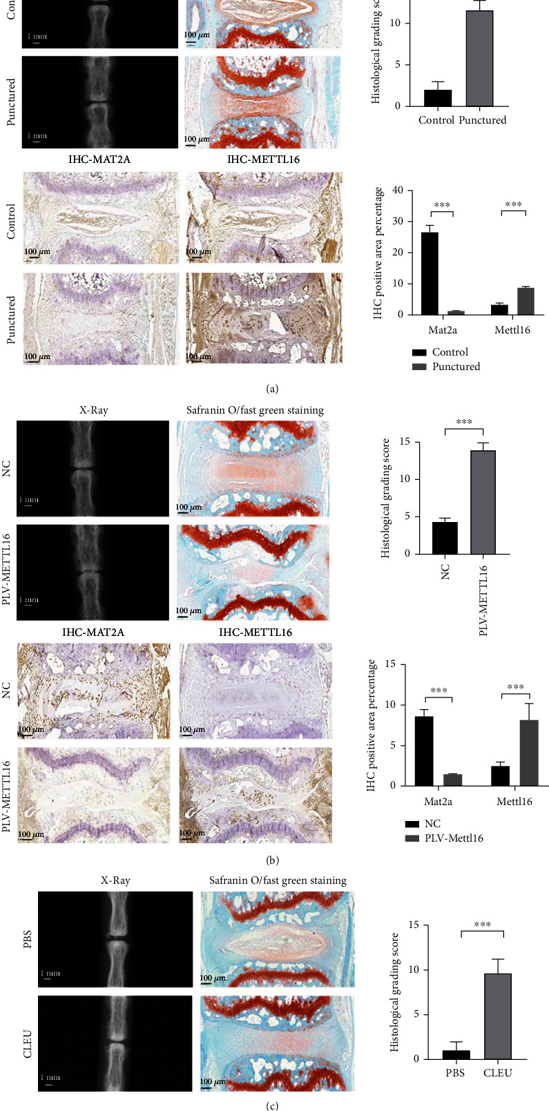
The process of intervertebral disc degeneration (IVDD) is complex, and its mechanism is considered multifactorial. Apoptosis of oxidative stressed nucleus pulposus cells (NPCs) should be a fundamental element in the pathogenesis of IVDD. In our pilot study, we found that the expression of MAT2A decreased, and METTL16 increased in the degenerative nucleus pulposus tissues. Previous studies have shown that the balance of splicing, maturation, and degradation of MAT2A pre-mRNA is regulated by METTL16 m6A modification. In the current study, we aimed to figure out whether this mechanism was involved in the aberrant apoptosis of NPCs and IVDD. Human NPCs were isolated and cultured under oxidative stress. An IVDD animal model was established. It showed that significantly higher METTL16 expression and lower MAT2A expression were seen in either the NPCs under oxidative stress or the degenerative discs of the animal model. MAT2A was inhibited with siRNA in vitro or cycloleucine in vivo. METTL16 was overexpressed with lentivirus in vitro or in vivo. Downregulation of MAT2A or upregulation of METTL16 aggravated nucleus pulposus cell apoptosis and disc disorganization. The balance of splicing, maturation, and degradation of MAT2A pre-mRNA was significantly inclined to degradation in the NPCs with the overexpression of METTL16. Increased apoptosis of NPCs under oxidative stress could be rescued by reducing the expression of METTL16 using siRNA with more maturation of MAT2A pre-mRNA. Collectively, oxidative stress aggravates apoptosis of NPCs through disrupting the balance of splicing, maturation, and degradation of MAT2A pre-mRNA, which is m6A modified by METTL16.


Conflict of interest statement

The authors declare that there is no conflict of interest regarding the publication of this paper.

Figures

Figure 1
Figure 1
MAT2A decreased and METTL16 increased in human degenerative nucleus pulposus. (a) The expression of MAT2A in human NP tissues was analyzed by qRT-PCR. (b) The expression of METTL16 in human NP tissues analyzed by qRT-PCR. (c) Immunofluorescence assay for the expression of MAT2A and METTL16 proteins in human NP tissues. ∗∗∗p < 0.001.
Figure 2
Figure 2
MAT2A was downregulated while METTL16 was upregulated when human nucleus pulposus cell was under oxidative stress. Human NPCs were exposed to TBHP (100 μm, 4 hours). (a) NPC identification by characterization of their cell surface marker CD24 (b) The expression of MAT2A mRNA was detected by qRT-PCR. (c) The expression of METTL16 mRNA was detected by qRT-PCR. (d) Western blotting for the expression of MAT2A and METTL16 proteins. (e) Immunofluorescence assay for the expression of MAT2A and METTL16 proteins. (f) SAM concentration in the cell culture medium supernatant was detected by ELISA. n = 3 replicates per group, ∗∗∗p < 0.001, 0.001 ≤  ∗∗p < 0.05, p < 0.05.
Figure 3
Figure 3
Downregulation of MAT2A or upregulation of METTL16 in NPCs promotes apoptosis. (a) Successful downregulation of MAT2A mRNA or upregulation of METTL16 mRNA in human NPCs confirmed by qRT-PCR. (b) SAM concentration in the supernatants of cell culture medium measured by ELISA method. (c) Apoptosis assayed by TUNEL staining significantly increased in the NPCs with MAT2A siRNA transfection. (d) Western blot analysis for the protein markers of apoptosis in cells with MAT2A siRNA transfection. (e) Flow cytometric analysis for the apoptosis of NPCs treated by MAT2A siRNA. (f) TUNEL staining for the apoptosis of NPCs with METTL16 overexpression. (g) The expression levels of Bcl2, Bax, and cleaved-Caspase3 detected by Western blot in cells with METTL16 overexpression. (h) Flow cytometric analysis of apoptosis in NPCs with METTL16 overexpression. n = 3 replicates per group, ∗∗∗p < 0.001, 0.001 ≤  ∗∗p < 0.05, p < 0.05.
Figure 4
Figure 4
The effect of METTL16 on MAT2A. (a) In degenerative human NP tissues, qRT-PCR showed that exon1-3 and harpin1 reflecting the total of MAT2A pre-mRNA and MAT2A mRNA decreased, while intron8 reflecting MAT2A pre-mRNA increased. (b) In the NPCs stimulated by TBHP, qRT-PCR showed exon1-3 and harpin1 decreased while intron8 increased. (c) In the NPCs with METTL16 overexpression, qRT-PCR showed MAT2A exon1-3 and harpin1 decreased, while intron8 increased. (d) Immunofluorescence showed that the expression of MAT2A protein decreased when METTL16 was upregulated in NPCs. The expression of MAT2A was higher in the NPCs with unsuccessful overexpression of METTL16 (white arrow) than in the cells with successful overexpression of METTL16 (black arrow). (e) MAT2A and METTL16 proteins detected by Western blot in cells with METTL16 overexpression. n = 3 replicates per group, ∗∗∗p < 0.001, 0.001 ≤  ∗∗p < 0.05, p < 0.05.
Figure 5
Figure 5
Increased apoptosis of human NPCs under oxidative stress can be rescued by reducing the expression of METTL16 in the cells. Human NPCs were transfected with METTL16 siRNA. (a) qRT-PCR analysis for transfection efficiency. (b) The cells were then stimulated with TBHP (100 μm, 4 hours). And qRT-PCR was employed to detect different fragments of MAT2A pre-mRNA. (c) MAT2A protein detected by immunofluorescence. (d) The protein expression levels of METTL16, MAT2A, Bcl2, Bax, and cleaved Caspase3 were detected by Western blot in cells with METTL16 siRNA and under TBHP stimulation. (e) Flow cytometry for the apoptosis rate of the NPCs. n = 3 replicates per group, ∗∗∗p < 0.001, 0.001 ≤  ∗∗p < 0.05, p < 0.05.
Figure 6
Figure 6
In vivo studies. (a) Verification of the disc degeneration animal model. X-ray examination revealed a significant decrease of the height of the punctured disc. Safranine O/fast green staining showed that the punctured NP tissue became more disorganized, and fewer cells could be seen. The histological grading score was significantly higher in the punctured disc. Immunohistochemical analysis demonstrated less MAT2A protein and more METTL16 protein levels in the punctured NP tissues. (b) Degenerative changes in the discs injected with METTL16 overexpression lentivirus. X-ray examination revealed significant decrease of the height of the discs. Safranine O/fast green staining showed that the NP tissue became more disorganized, and fewer cells could be seen. The histological grading score was also significantly higher. Immunohistochemical analysis demonstrated less MAT2A protein and more METTL16 protein levels in the NP tissues. (c) Degenerative changes in the discs injected with cycloleucine. X-ray examination revealed significant decrease of the height of the discs. Safranine O/fast green staining showed that the NP tissue became more disorganized, and fewer cells could be seen. The histological grading score was also significantly higher. n = 3 replicates per group, ∗∗∗p < 0.001, 0.001 ≤  ∗∗p < 0.05, p < 0.05.

References

    1. Bressler H. B., Keyes W. J., Rochon P. A., Badley E. The prevalence of low back pain in the Elderly. Spine . 1999;24(17):1813–1819. doi: 10.1097/00007632-199909010-00011. - DOI - PubMed
    1. Hoy D., Bain C., Williams G., et al. A systematic review of the global prevalence of low back pain. Arthritis and Rheumatism . 2012;64(6):2028–2037. doi: 10.1002/art.34347. - DOI - PubMed
    1. Prince M. J., Wu F., Guo Y., et al. The burden of disease in older people and implications for health policy and practice. Lancet (London, England) . 2015;385(9967):549–562. doi: 10.1016/S0140-6736(14)61347-7. - DOI - PubMed
    1. Roberts S., Evans H., Trivedi J., Menage J. Histology and pathology of the human intervertebral disc. The Journal of bone and joint surgery American . 2006;88(supplement 2):10–14. doi: 10.2106/JBJS.F.00019. - DOI - PubMed
    1. Zhao C. Q., Wang L. M., Jiang L. S., Dai L. Y. The cell biology of intervertebral disc aging and degeneration. Ageing Research Reviews . 2007;6(3):247–261. doi: 10.1016/j.arr.2007.08.001. - DOI - PubMed

LinkOut - more resources