Review
. 2021 Dec 9:12:50.
doi: 10.4103/jpi.jpi_66_21. eCollection 2021.

Contemporary Whole Slide Imaging Devices and Their Applications within the Modern Pathology Department: A Selected Hardware Review

Affiliations
Review

Contemporary Whole Slide Imaging Devices and Their Applications within the Modern Pathology Department: A Selected Hardware Review

Ankush Patel et al. J Pathol Inform. .

Abstract

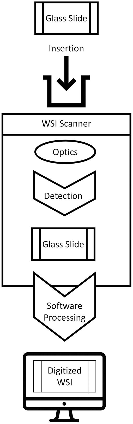
Digital pathology (DP) has disrupted the practice of traditional pathology, including applications in education, research, and clinical practice. Contemporary whole slide imaging (WSI) devices include technological advances that help address some of the challenges facing modern pathology, such as increasing workloads with fewer subspecialized pathologists, expanding integrated delivery networks with global reach, and greater customization when working up cases for precision medicine. This review focuses on integral hardware components of 43 market available and soon-to-be released digital WSI devices utilized throughout the world. Components such as objective lens type and magnification, scanning camera, illumination, and slide capacity were evaluated with respect to scan time, throughput, accuracy of scanning, and image quality. This analysis of assorted modern WSI devices offers essential, valuable information for successfully selecting and implementing a digital WSI solution for any given pathology practice.

Keywords: Digital pathology; hardware; microscopy; slide scanning; whole slide imaging; whole slide scanner.


Conflict of interest statement

There are no conflicts of interest.

Figures

Figure 1
Figure 1
WSI creation (original illustration)
Figure 2
Figure 2
Stage movement and image capture (original illustration, based on 3D Histech Pannoramic 250 Flash III camera and stage). Glass slide with specimen (pink) driven through the WSI scanner via continuous movement of the motorized stage. A lens objective is shown above the glass slide. Continuous stage movement is accompanied by rapid, repetitive image capture via a camera depicted below the glass slide. The 3D Histech Pannoramic 250 Flash III has an image capture rate of 130 frames per second[35]
Figure 3
Figure 3
WSI device exterior and interior (original illustration based on 3D Histech Pannoramic 250 Flash III Configuration). WSI Device Exterior (A) and interior (B, C) components consisting of multiple objectives along with fluorescence and brightfield capabilities (Modified from 3D Histech Pannoramic 250 Flash III).[35]
Figure 4
Figure 4
Field of view (original illustration). (A) Maximal field of view elicited by the lens objective. (B) Superimposed sensor FOV. (C) FOV demarking image area seen by viewer
Figure 5
Figure 5
Scanning methods: line vs. tile (original illustration). (A)Tile/area scanning utilizing focus points in every field. (B) Tile/ area scanning utilizing focus in every “nth” field”. (C) Line scanning utilizing “focus map” technique in which focus points dictate direction of scanning (indicated by yellow arrows). (D) Image “stitching” after digital data acquisition
Figure 6
Figure 6
“Z-Stack/ extended depth of field” (original illustration). “Z-stacking,” a method of scanning by which a series of images are captured at various focal planes, i.e., “slices,” that are then combined to most effectively portray samples with 3D structures, such as clumps of cells or thick tissue. Some WSI scanning methods are built upon the Z-stack technique, e.g., “Extended Depth of Field (EDF),” or extended focus, which further enhances Z-stacking by combining the sharpest points of focus from each slice to maximize the depth of sharpness in the final image, demonstrated to be useful in cytology applications.
Figure 7
Figure 7
Extended focus (figure is not an original illustration and has been sourced from a 3D Histech company presentation). (A and B) Application of extended focus courtesy of 3D Histech

References

    1. Browning L, Colling R, Rakha E, Rajpoot N, Rittscher J, James JA, et al. Digital pathology and artificial intelligence will be key to supporting clinical and academic cellular pathology through COVID-19 and future crises: The PathLAKE consortium perspective. J Clin Pathol. 2021;74:443–7. - PMC - PubMed
    1. Evans AJ, Depeiza N, Allen SG, Fraser K, Shirley S, Chetty R. Use of whole slide imaging (WSI) for distance teaching. J Clin Pathol. 2021;74:425–8. - PubMed
    1. Montironi R, Cimadamore A, Scarpelli M, Cheng L, Lopez-Beltran A, Mikuz G. Pathology without microscope: From a projection screen to a virtual slide. Pathol Res Pract. 2020;216:153196. - PMC - PubMed
    1. Pallua JD, Brunner A, Zelger B, Schirmer M, Haybaeck J. The future of pathology is digital. Pathol Res Pract. 2020;216:153040. - PubMed
    1. Weinstein RS. Prospects for telepathology. Hum Pathol. 1986;17:433–4. - PubMed

LinkOut - more resources