. 2022 Jan 7:11:789398.
doi: 10.3389/fcimb.2021.789398. eCollection 2021.

TNF-TNFR1 Signaling Enhances the Protection Against Neospora caninum Infection

Affiliations

TNF-TNFR1 Signaling Enhances the Protection Against Neospora caninum Infection

Flávia Batista Ferreira França et al. Front Cell Infect Microbiol. .

Abstract

Neospora caninum is a protozoan associated with abortions in ruminants and neuromuscular disease in dogs. Classically, the immune response against apicomplexan parasites is characterized by the production of proinflammatory cytokines, such as IL-12, IFN-γ and TNF. TNF is mainly produced during the acute phases of the infections and binds to TNF receptor 1 (CD120a, p55, TNFR1) activating a variety of cells, hence playing an important role in the induction of the inflammatory process against diverse pathogens. Thus, in this study, we aimed to evaluate the role of TNF in cellular and humoral immune responses during N. caninum infection. For this purpose, we used a mouse model of infection based on wildtype (WT) and genetically deficient C57BL/6 mice in TNFR1 (Tnfr1-/-). We observed that Tnfr1-/- mice presented higher mortality associated with inflammatory lesions and increased parasite burden in the brain after the infection with N. caninum tachyzoites. Moreover, Tnfr1-/- mice showed a reduction in nitric oxide (NO) levels in vivo. We also observed that Tnfr1-/- mice showed enhanced serum concentration of antigen-specific IgG2 subclass, while IgG1 production was significantly reduced compared to WT mice, suggesting that TNFR1 is required for regular IgG subclass production and antigen recognition. Based on our results, we conclude that the TNF-TNFR1 complex is crucial for mediating host resistance during the infection by N. caninum.

Keywords: TNF; antibodies; chronic phase; effector molecules; neosporosis.


Conflict of interest statement

The authors declare that the research was conducted in the absence of any commercial or financial relationships that could be construed as a potential conflict of interest.

Figures

Figure 1
Figure 1
TNF is crucial for the survival of mice against Neospora caninum. Groups of WT and Tnfr1 -/- mice were infected with 1×10⁷ Nc-1 tachyzoites and survival was observed for 30 days. Differences between groups were compared using Kaplan Meier survival analysis, through the log-rank (Mantel-Cox) test. Statistically significant differences (p < 0.05). Two independent experiments were performed with similar results, and the graphs represent the biological replicates (n = 10 mice/group) of one of those individual experiments.
Figure 2
Figure 2
TNF is required to control the parasite burden during the chronic phase of infection. WT and Tnfr1-/- mice (n = 5 mice/group) infected with 1×10⁶ Nc-1 tachyzoites were evaluated for parasite burden by qPCR. Peritoneal exudates (A), lungs (B), liver (C) and brains (D) were evaluated for the number of copies of the genomic sequence NC5. Two independent experiments were performed with similar results, and the graphs represent the biological replicates (n = 5 mice/group) of one of those individual experiments. Results are expressed as mean ± standard error of the mean (SEM). Data were analyzed using the Mann Whitney test. *Statistically significant differences (p < 0.05).
Figure 3
Figure 3
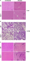
TNFR1 controls cerebral inflammation during chronification of N. caninum infection. Representative histological images of hepatic (A) and pulmonary (B) tissue sections analyzed 7 days post-infection, and brain sections (C) - collected at 30 days post-infection - in WT and Tnfr1 -/- mice infected with 1 × 10⁶ tachyzoites of N. caninum. The histological sections were stained with H&E and analyzed under an optical microscope. Some focal inflammatory infiltrates are indicated by black arrows. Scale bar: 150µm. Two independent experiments were performed with similar results, and each figure represents the tissue of one biological replicate of one of those individual experiments. Tissues were obtained from 5 mice/group in each experiment.
Figure 4
Figure 4
While TNFR1 does not downmodulate the production of key Th1 cytokines during the infection, it is essential for the production of proper levels of nitric oxide (NO). Groups of WT and Tnfr1 -/- mice were evaluated for IL-12p40 (A) and IFN-γ (B) production 7 days after the infection with 1×10⁶ Nc-1 tachyzoites in the peritoneal fluids, serum samples, lung and liver homogenates. NO concentration was evaluated 7 days after infection in peritoneal fluids (C). Values are expressed as mean ± standard error of the mean (SEM). Data were analyzed using the Two-way ANOVA test, followed by Bonferroni post-test. Differences were considered statistically significant (*) when p < 0.05. For the in vivo assays, two independent experiments were performed with similar results, and the graphs represent the biological replicates (n = 5 mice/group) of one of those individual experiments.
Figure 5
Figure 5
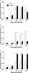
TNFR1 controls the production of IgG subclasses against soluble antigens. WT and Tnfr1 -/- mice were infected intraperitoneally with 1×10⁶ Nc-1 tachyzoites and sampled weekly for serum samples until 28 days of infection. Levels of IgG (A), IgG1 (B) and IgG2a (C) were determined by ELISA and values are indicated as optical density (OD) at 405nm, being expressed as mean ± standard error of the mean (SEM). Data were analyzed using the Two-way ANOVA test followed by Bonferroni post-test. Differences were considered statistically significant (*) when p < 0.05. Two independent experiments were performed with similar results, and the graphs represent the biological replicates (n = 5 mice/group) of one of those individual experiments.
Figure 6
Figure 6
Immunoblots show the lack of proper antigen recognition in the absence of TNFR1. Individual serum samples of WT and Tnfr1 -/- mice, obtained at 0, 14 and 28 days after the infection by Neospora caninum, were submitted to immunoblots for the determination of the pattern of recognition by specific IgG (A), IgG1 (B) and IgG2a (C) antibodies against the parasite’s soluble antigen. We have displayed immunoblots that are representative of the reactivity obtained by an individual mouse/group/antibody, and that had similar results in two independent experiments with 5 mice/group each.

References

    1. Aggarwal B. B., Gupta S. C., Kim J. H. (2012). Historical Perspectives on Tumor Necrosis Factor and its Superfamily: 25 Years Later, A Golden Journey. Blood 119, 651–665. doi: 10.1182/blood-2011-04-325225 - DOI - PMC - PubMed
    1. Aguado-Martinez A., Basto A. P., Leitao A., Hemphill A. (2017). Neospora Caninum in Non-Pregnant and Pregnant Mouse Models: Cross-Talk Between Infection and Immunity. Int. J. Parasitol. 47, 723–735. doi: 10.1016/j.ijpara.2017.09.001 - DOI - PubMed
    1. Almeria S., Serrano-Perez B., Lopez-Gatius F. (2017). Immune Response in Bovine Neosporosis: Protection or Contribution to the Pathogenesis of Abortion. Microb. Pathog. 109, 177–182. doi: 10.1016/j.micpath.2017.05.042 - DOI - PubMed
    1. Barros P. D. S. C., Mota C. M., dos Santos Miranda V., Ferreira F. B., Ramos E. L. P., Santana S. S., et al. . (2019). Inducible Nitric Oxide Synthase Is Required for Parasite Restriction and Inflammatory Modulation During Neospora Caninum Infection. Vet. Parasitol. 276, 108990. doi: 10.1016/j.vetpar.2019.108990 - DOI - PubMed
    1. Bjerkas I., Mohn S. F., Presthus J. (1984). Unidentified Cyst-Forming Sporozoon Causing Encephalomyelitis and Myositis in Dogs. Z. Parasitenkd. 70, 271–274. doi: 10.1007/BF00942230 - DOI - PubMed

Publication types

Substances

LinkOut - more resources