. 2022 Jan 5:9:750878.
doi: 10.3389/fcell.2021.750878. eCollection 2021.

Exogenous PTH 1-34 Attenuates Impaired Fracture Healing in Endogenous PTH Deficiency Mice via Activating Indian Hedgehog Signaling Pathway and Accelerating Endochondral Ossification

Affiliations

Exogenous PTH 1-34 Attenuates Impaired Fracture Healing in Endogenous PTH Deficiency Mice via Activating Indian Hedgehog Signaling Pathway and Accelerating Endochondral Ossification

Cheng Ma et al. Front Cell Dev Biol. .

Abstract

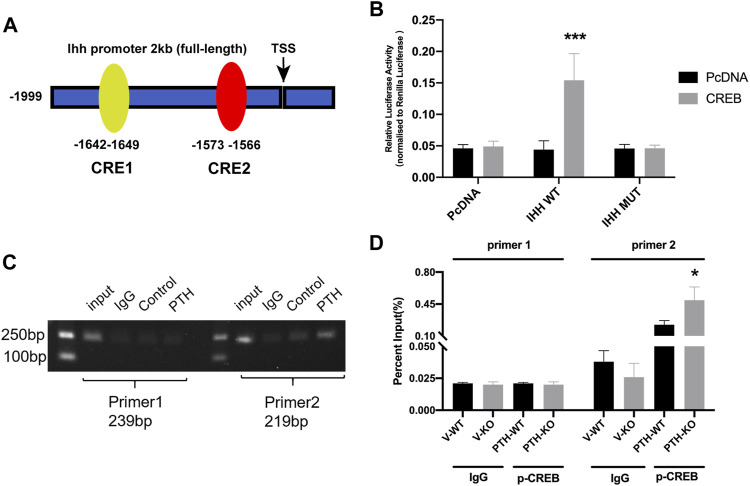
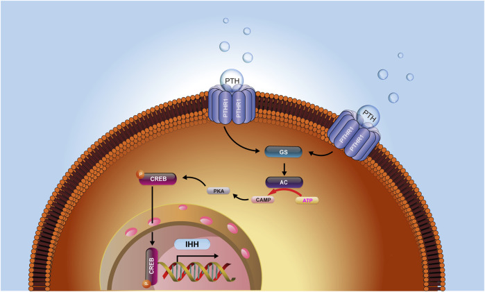
Fracture healing is a complicated, long-term, and multistage repair process. Intermittent administration of parathyroid hormone (PTH) has been proven effective on intramembranous and endochondral bone formation during the fracture healing process, however, the mechanism is unclear. In this study, we investigated the role of exogenous PTH and endogenous PTH deficiency in bone fracture healing and explored the mechanism by using PTH knockout (PTH-/-) mice and ATDC5 cells. In a mouse femur fracture model, endogenous PTH deficiency could delay endochondral ossification whereas exogenous PTH promotes accumulation of endochondral bone, accelerates cartilaginous callus conversion to bony callus, enhances maturity of bony callus, and attenuates impaired fracture healing resulting from endogenous PTH deficiency. In fracture callus tissue, endogenous PTH deficiency could inhibit chondrocyte proliferation and differentiation whereas exogenous PTH could activate the IHH signaling pathway to accelerate endochondral ossification and rescue impaired fracture healing resulting from endogenous PTH deficiency. In vitro, exogenous PTH promotes cell proliferation by activating IHH signaling pathway on ATDC5 cells. In mechanistic studies, by using ChIP and luciferase reporter assays, we showed that PTH could phosphorylate CREB, and subsequently bind to the promoter of IHH, causing the activation of IHH gene expression. Therefore, results from this study support the concept that exogenous PTH 1-34 attenuates impaired fracture healing in endogenous PTH deficiency mice via activating the IHH pathway and accelerating endochondral ossification. Hence, the investigation of the mechanism underlying the effects of PTH treatment on fracture repair might guide the exploration of effective therapeutic targets for fracture.

Keywords: Indian hedgehog signaling pathway; chondrocyte; endochondral ossification; fracture healing; parathyroid hormone.


Conflict of interest statement

The authors declare that the research was conducted in the absence of any commercial or financial relationships that could be construed as a potential conflict of interest.

Figures

FIGURE 1
FIGURE 1
The role of endogenous PTH deficiency and exogenous PTH on cartilaginous and bony callus formation. (A) Representative images of H&E staining of paraffin callus sections from V-WT, V-KO, PTH-WT, and PTH-KO groups at 7, 14, 21 days post-fracture, Scale bar: 50 μm. (B) Representative images of Safranin-O/fast green staining of paraffin callus sections from V-WT, V-KO, PTH-WT, and PTH-KO groups at 7, 14, 21 days post-fracture, Scale bar: 50 μm. (C) Representative radiographs of fractured femurs from V-WT, V-KO, PTH-WT, and PTH-KO groups at 7, 14, 21, and 28 days post-fracture. (D) Quantitative assessment of cartilaginous callus area, bony callus, and total callus area of V-WT, V-KO, PTH-WT, and PTH-KO groups at 7 and 14 days post-fracture. (E,F) Representative longitudinal micro-CT and 3D reconstructions images of the fractured femurs from V-WT, V-KO, PTH-WT, and PTH-KO groups at 7, 14, 21, and 28 days post-fracture. Quantitative assessment of fracture callus of V-WT, V-KO, PTH-WT, and PTH-KO groups at 14 days post-fracture. (G) callus volume (H) bone mineral density (I) bone volume/total volume (J) trabecular number (K) trabecular thickness (L) trabecular separation. All data are expressed as mean ± SD (n = 3). (*p < 0.05; **p < 0.01; ***p < 0.001; compared with control group. # p < 0.05; ## p < 0.01; ### p < 0.001; compared with V-KO group).
FIGURE 2
FIGURE 2
The role of endogenous PTH deficiency and exogenous PTH on chondrocyte proliferation and differentiation at the callus. (A) The qRT-PCR analysis of Col Ⅱ, Col X, PTHR1 and PCNA mRNA expression in V-WT, V-KO, PTH-WT, and PTH-KO group at 7 and 14 days post-fracture. GAPDH was used for normalization (n = 3/group). (B,C) Western blot analysis of the IHH, SMO, Gli1, and Gli3 protein expression in V-WT, V-KO, PTH-WT, and PTH-KO group at 7 and 14 days post-fracture and the p-CREB and CREB protein expression at 7 days post-fracture. β-Actin was used for normalization (n = 3/group). (D) Representative images of IHC staining of paraffin callus sections for Col Ⅱ, Col X, PTHR1, p-CREB, and PCNA from V-WT, V-KO, PTH-WT, and PTH-KO mice at 7 and 14 days post-fracture, Scale bar: 25 μm. (E) Quantitative analysis of IHC staining. All data are expressed as mean ± SD (n = 3). (*p < 0.05; **p < 0.01; ***p < 0.001; compared with control group. # p < 0.05; ## p < 0.01; ### p < 0.001; compared with V-KO group).
FIGURE 3
FIGURE 3
The role of endogenous PTH deficiency and exogenous PTH on the regulation of Indian hedgehog signaling pathway at the callus. (A) The qRT-PCR analysis of IHH, SMO, Gli1, and Gli3 mRNA expression in V-WT, V-KO, PTH-WT, and PTH-KO groups at 7 and 14 days post-fracture. GAPDH was used for normalization (n = 3/group). (B,C) Western blot analysis of the IHH, SMO, and Gli1 protein expression in V-WT, V-KO, PTH-WT, and PTH-KO groups at 7 and 14 days post-fracture. β-Actin was used for normalization (n = 3/group). (D) Representative images of IHC staining of paraffin callus sections for IHH, SMO, Gli1, and Gli3 from V-WT, V-KO, PTH-WT, and PTH-KO groups at 7 and 14 days post-fracture, Scale bar: 25 μm. (E) Quantitative analysis of IHC staining. All data are expressed as mean ± SD (n = 3). (*p < 0.05; **p < 0.01; ***p < 0.001; compared with control group. # p < 0.05; ## p < 0.01; ### p < 0.001; compared with V-KO group).
FIGURE 4
FIGURE 4
The exogenous PTH promotes proliferation by activating IHH signaling pathway on ATDC5 cells. (A) Growth curves of ATDC5 cells at different concentrations of PTH1-34 measured by CCK8-assay (n = 6/group) (*p < 0.05, 0 mol/L vs. 10−7 mol/L, # p < 0.05, 0 mol/L vs. 10−8 mol/L, & p < 0.05, 0 mol/L vs. 10−9 mol/L, ^ p < 0.05, 0 mol/L vs. 10−10 mol/L). (B) Toluidine blue staining and Alcian blue staining in ATDC5 cells at days 7 after incubation with different concentrations of PTH1-34, Scale bar: 50 μm. (C) The qRT-PCR analysis of Col Ⅱ, Col X, PCNA, PTHR1, IHH, SMO, Gli1, and Gli3 mRNA expression in the groups at day 4 in the presence or absence of PTH (10−7 mol/L) and cyclopamine (10−6 mol/L). GAPDH was used for normalization (n = 3/group). (D,E) Western blot analysis of the Col Ⅱ, Col X, PTHR1, p-CREB, CREB, IHH, SMO, Gli1, and Gli3 protein expression in the groups at day 4 in the presence or absence of PTH (10−7 mol/L) and cyclopamine (10−6 mol/L). β-Actin was used for normalization (n = 3/group). (F) Representative IF staining images of Col Ⅱ, Col X, PCNA, PTHR1, p-CREB, IHH, Smo, Gli1, and Gli3 expression levels in ATDC5 cells in the groups at days 4 in the presence or absence of PTH (10−7 mol/L) and cyclopamine (10−6 mol/L), Scale bar: 20 μm. (G) Quantitative analysis of IF staining. All data are expressed as mean ± SD (n = 3). (*p < 0.05; **p < 0.01; ***p < 0.001; compared with control group. # p < 0.05; ## p < 0.01; ### p < 0.001; compared with PTH group).
FIGURE 5
FIGURE 5
The transcription factor CREB facilitates IHH expression through promoter activation. (A) A diagram shows the relative positions of full-length (FL) and fragments of IHH promoter reporters. (B) The dual-luciferase assays show that overexpression of CREB significantly increased the luciferase activity of WT-IHH but not MUT-IHH. (C,D) ChIP-qPCR shows that PTH could phosphorylate CREB and directly bind to −1642 to −1649 bp of IHH promoter. All data are expressed as mean ± SD (n = 3). (*p < 0.05; **p < 0.01; ***p < 0.001, compared with control group).
FIGURE 6
FIGURE 6
The hypothesis model depicts that PTH could increase intracellular cAMP concentrations and activate the signaling transduction of the cAMP/PKA/CREB axis, causing a subsequent increase of the level of phosphorylated CREB, which in turn activated the IHH signaling pathway. IHH further led to increased chondrogenesis in the callus and an enhanced rate of chondrocyte maturation and mineralization and ultimately accelerated the process of fracture healing.

References

    1. Alkhiary Y. M., Gerstenfeld L. C., Krall E., Westmore M., Sato M., Mitlak B. H., et al. (2005). Enhancement of Experimental Fracture-Healing by Systemic Administration of Recombinant Human Parathyroid Hormone (PTH 1-34). J. Bone Jt. Surg. 87 (4), 731–741. 10.2106/JBJS.D.02115 - DOI - PubMed
    1. Andreassen T. T., Ejersted C., Oxlund H. (1999). Intermittent Parathyroid Hormone (1-34) Treatment Increases Callus Formation and Mechanical Strength of Healing Rat Fractures. J. Bone Miner Res. 14 (6), 960–968. 10.1359/jbmr.1999.14.6.960 - DOI - PubMed
    1. Chang J.-K., Chang L.-H., Hung S.-H., Wu S.-C., Lee H.-Y., Lin Y.-S., et al. (2009). Parathyroid Hormone 1-34 Inhibits Terminal Differentiation of Human Articular Chondrocytes and Osteoarthritis Progression in Rats. Arthritis Rheum. 60 (10), 3049–3060. 10.1002/art.24843 - DOI - PubMed
    1. Chen M. Q., Luan J. J. (2019). HMGB1 Promotes Bone Fracture Healing through Activation of ERK Signaling Pathway in a Rat Tibial Fracture Model. Kaohsiung J. Med. Sci. 35 (9), 550–558. 10.1002/kjm2.12095 - DOI - PMC - PubMed
    1. de Crombrugghe B., Lefebvre V., Nakashima K. (2001). Regulatory Mechanisms in the Pathways of Cartilage and Bone Formation. Curr. Opin. Cel Biol. 13 (6), 721–728. 10.1016/s0955-0674(00)00276-3 - DOI - PubMed

LinkOut - more resources