Retroperitoneal parasitic fetus: A case report
- PMID: 35071581
- PMCID: PMC8717500
- DOI: 10.12998/wjcc.v9.i36.11482
Retroperitoneal parasitic fetus: A case report
Abstract
Background: Fetus-in-fetu (FIF) is an extremely rare congenital abnormal mass, in which a normal fetus's vertebral axis frequently connected with malformed fetus around this axis. Here, we report the case of a male infant aged 26 d presenting with retroperitoneal parasitic fetus.
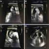
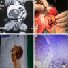
Case summary: In a prenatal examination, we first detected an abdominal mass measuring 7.8 cm × 5.1 cm × 6.8 cm in a mother's abdomen at 25 gestational weeks and teratoma was suspected. After the fetal was born, we did a magnetic resonance imaging (MRI) and ultrasonography on him and saw a distinctive limb with five-toes. According to the result of MRI, ultrasonography and postoperative pathology, he finally was diagnosed with FIF.
Conclusion: A laparotomy was performed at 26 d of age with excision of the retroperitoneal cystic tumor, which measured about 10 cm in diameter. According to the result of imaging and histological test, FIF was confirmed.
Keywords: Abdominal mass; Case report; Fetus-in-fetu; Teratoma; Ultrasound.
©The Author(s) 2021. Published by Baishideng Publishing Group Inc. All rights reserved.
Conflict of interest statement
Conflict-of-interest statement: The authors declare that there is no conflict of interest.
Figures


References
-
- Grant P, Pearn JH. Foetus-in-foetu. Med J Aust. 1969;1:1016–1019. - PubMed
-
- Sathe PA, Ghodke RK, Kandalkar BM. Fetus in fetu: an institutional experience. Pediatr Dev Pathol. 2014;17:243–249. - PubMed
-
- Ruffo G, Di Meglio L, Sica C, Resta A, Cicatiello R. Fetus-in-fetu: two case reports. J Matern Fetal Neonatal Med. 2019;32:2812–2819. - PubMed
-
- Sharifah MI, Noryati M, Che Zubaidah CD, Zakaria Z. Foetus-in-Fetu. Med J Malaysia. 2010;65:150–151. - PubMed
Publication types
LinkOut - more resources
Full Text Sources

