. 2022 Jan 11;3(1):101084.
doi: 10.1016/j.xpro.2021.101084. eCollection 2022 Mar 18.

Protocol for the assessment of human T cell activation by real-time metabolic flux analysis

Affiliations

Protocol for the assessment of human T cell activation by real-time metabolic flux analysis

Byung Soo Kong et al. STAR Protoc. .

Abstract

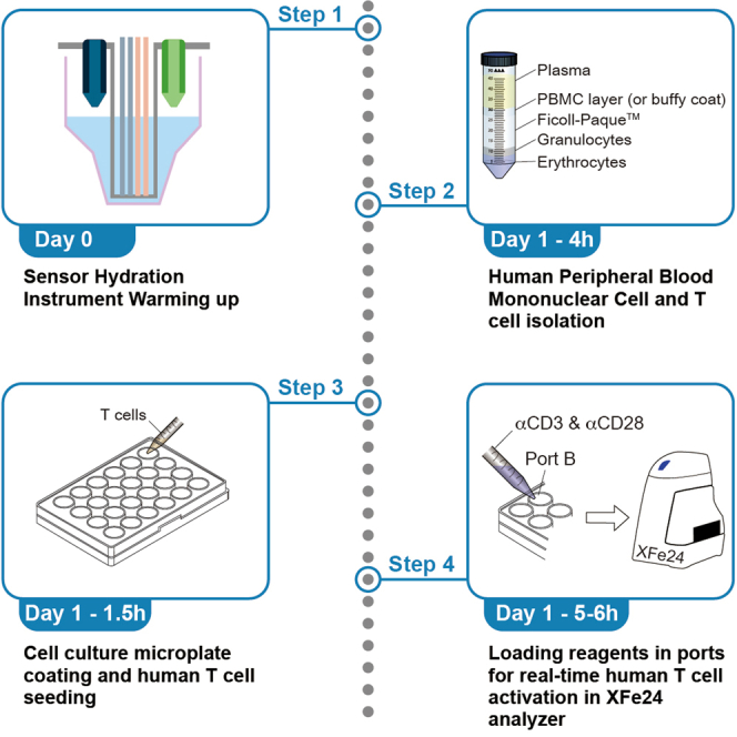
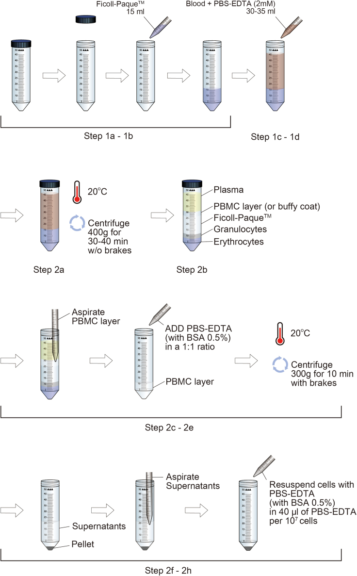
The elevation of glycolysis in autoreactive T cells is a key target for the prevention and treatment of T cell-related autoimmune diseases, such as type 1 diabetes (T1D). Here, we describe a simple and efficient protocol for isolating human peripheral blood mononuclear cells (PBMCs) and T cells, and the subsequent assessment of T cell glycolysis using Seahorse analyzer. This protocol is useful to analyze different subsets of T cells and applicable to different autoimmune disease models (i.e., T1D, multiple sclerosis). For complete details on the use and execution of this profile, please refer to Kong et al. (2021).

Keywords: Cell isolation; Cell-based Assays; Classification Description: Cell Biology; Immunology; Metabolism.


Conflict of interest statement

The authors declare no competing interests.

Figures

None
Graphical abstract
Figure 1
Figure 1
Isolation of peripheral blood mononuclear cells (PBMCs) using density gradient centrifugation This protocol is optimized for the preparation of PBMCs from human blood using Ficoll-paqueTM. This figure is to show detailed steps for “before you begin”
Figure 2
Figure 2
Preparation of XF running medium and XF cell culture microplate for human T cells This figure is to show detailed steps for “step-by-step method details”. pH and temperature of XF running medium is important factor in the experiment. Also, coating cell culture microplate for human T cells is essential to allow cells to be positioned at the right spot for the measurement by sensor cartridge.
Figure 3
Figure 3
Loading ports in the microplate cartridge for drug and soluble αCD3 and αCD28 injections This figure is to show detailed steps for “step-by-step method details”. The schematic diagram of XFe24 cell culture microplate shows that the wells are double-layered in the top view. The side view of XFe24 cell culture microplate also shows that cells and medium should be added separately to avoid cells seeding on the top-level. The XFe24 Sensor Cartridge diagram has to be hydrated in utility plate before use. Also, the compounds and soluble αCD3 and αCD28 are loaded in this component as shown in steps 8 and 9.
Figure 4
Figure 4
Diagram showing the Wave program set up for real-time T cell activation assessment This figure is to show detailed steps for “step-by-step method details”. Designing the program for the experiment is shown here. The program set-up can be done before the experiment and the template can be saved for future analyses.
Figure 5
Figure 5
Successful (left) and failed (right) outcome after performing metabolic flux analysis in human T cells (A) The successful outcome shows that the ECAR level of human T cells after the treatment of soluble αCD3 and αCD28 is increased from 2 to 4 mpH/min. Data are presented as mean ± standard error of mean (SEM) (B) However, the failed outcome shows that the treatment of soluble αCD3 and αCD28 did not increase the ECAR level of human T cells. Data are presented as mean ± standard error of mean (SEM)

References

    1. Ahn C.H., Choi E.H., Kong B.S., Cho Y.M. Effects of MOTS-c on the mitochondrial function of cells harboring 3243 A to G mutant mitochondrial DNA. Molecular Biology Reports. 2020;47:4029–4035. - PubMed
    1. Boyum A. Isolation of lymphocytes, granulocytes and macrophages. Scand. J. Immunol. 1976;Suppl 5:9–15. - PubMed
    1. Buhl T., Legler T.J., Rosenberger A., Schardt A., Schon M.P., Haenssle H.A. Controlled-rate freezer cryopreservation of highly concentrated peripheral blood mononuclear cells results in higher cell yields and superior autologous T-cell stimulation for dendritic cell-based immunotherapy. Cancer Immunol. Immunother. 2012;61:2021–2031. - PMC - PubMed
    1. Cedrone E., Neun B.W., Rodriguez J., Vermilya A., Clogston J.D., McNeil S.E., Barenholz Y., Szebeni J., Dobrovolskaia M.A. Anticoagulants influence the performance of in vitro assays intended for characterization of nanotechnology-based formulations. Molecules. 2017;23:12. - PMC - PubMed
    1. Diks A.M., Bonroy C., Teodosio C., Groenland R.J., de Mooij B., de Maertelaere E., Neirynck J., Philippe J., Orfao A., van Dongen J.J.M., et al. Impact of blood storage and sample handling on quality of high dimensional flow cytometric data in multicenter clinical research. J. Immunol. Methods. 2019;475:112616. - PubMed

Publication types