. 2022 Mar;9(9):e2105477.
doi: 10.1002/advs.202105477. Epub 2022 Jan 24.

Application of Raman Spectroscopy for Sorption Analysis of Functionalized Porous Materials

Affiliations

Application of Raman Spectroscopy for Sorption Analysis of Functionalized Porous Materials

Gregor Lipinski et al. Adv Sci (Weinh). 2022 Mar.

Abstract

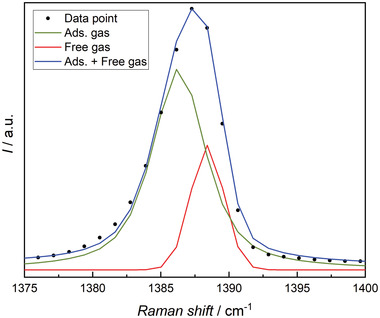
Functionalized porous materials could play a key role in improving the efficiency of gas separation processes as required by applications such as carbon capture and storage (CCS) and across the hydrogen value chain. Due to the large number of different functionalizations, new experimental approaches are needed to determine if an adsorbent is suitable for a specific separation task. Here, it is shown for the first time that Raman spectroscopy is an efficient tool to characterize the adsorption capacity and selectivity of translucent functionalized porous materials at high pressures, whereby translucence is the precondition to study mass transport inside of a material. As a proof of function, the performance of three silica ionogels to separate an equimolar (hydrogen + carbon dioxide) gas mixture is determined by both accurate gravimetric sorption measurements and Raman spectroscopy, with the observed consistency establishing the latter as a novel measurement technique for the determination of adsorption capacity. These results encourage the use of the spectroscopic approach as a rapid screening method for translucent porous materials, particularly since only very small amounts of sample are required.

Keywords: Raman spectroscopy; adsorption; functionalized porous materials; gas separation.


Conflict of interest statement

The authors declare no conflict of interest.

Figures

Figure 1
Figure 1
Schematic of the experimental setup for adsorption measurements of an equimolar gas mixture on an IG utilizing a confocal Raman microscope. Spectra are recorded along a single axis at equidistant intervals of 5 µm in the bulk phase (right in the optical cell) and inside the IG particle (left in the optical cell). [We note that this interval was chosen to ensure that there is no overlap between the measured points. As the measurements take place well inside the particle, overlapping points might not be an issue so other intervals would be possible without affecting the result.] Measurements are conducted at z = 200 µm, which corresponds to approximately half the height of the investigated particles. The structure of a mesopore is highlighted in the inset. Please note: The details in the schematic are not true to scale.
Figure 2
Figure 2
Partial Raman spectra used for the analysis of the adsorption of carbon dioxide, measured for the vapor phase (red) and in IG1 (black) at T = 293.15 K and p = 50 bar. The main peak can be de‐convolved into free and adsorbed gas peaks as shown in Figure 6.
Figure 3
Figure 3
Mapped concentration of carbon dioxide (top) and hydrogen (bottom) on IG1 and photograph of the respective ionogel particle (left). Measurements were conducted at T = 293.15 K and p = (20, 30, 40, 50) bar. Brighter pixel is assigned to a higher concentration of carbon dioxide and hydrogen, respectively. Each pixel has a size of 2,25 µm2.
Figure 4
Figure 4
Results of spectroscopic and gravimetric adsorption measurements of pure carbon dioxide on IG1 (red), IG2 (green), and IG3 (blue) along the 293.15 K isotherm. Raman data are shown with filled symbols and gravimetric data with empty symbols. Error bars for the expanded uncertainty of net adsorption U C(qnet) with (k = 2) are included for every measured (T, p) state point. Additional lines are plotted to guide the eye.
Figure 5
Figure 5
Results of spectroscopic and gravimetric adsorption measurements of an equimolar (hydrogen + carbon dioxide) gas mixture on IG1 (red), IG2 (green), and IG3 (blue) along the 293.15 K isotherm. Results are for carbon dioxide adsorption capacity. Raman data are shown with filled symbols and gravimetric data with empty symbols. Error bars for the expanded combined uncertainty (k = 2) of net adsorption U C(qnet) are included for every measured (T, p) state point. Additional lines are plotted to guide the eye.
Figure 6
Figure 6
Schematic of a deconvolution analysis. The signal of the Fermi dyad recorded at T = 293.15 K and p = 50 bar in a range of (1375–1400) cm−1 is deconvolved into two separate signals that are assigned to free (red) and adsorbed (green) carbon dioxide molecules. The original Raman data points (black dots) and the sum of the two fitted peaks (blue) are also shown.

References

    1. Welton T., Chem. Rev. 1999, 99, 2071. - PubMed
    1. Hapiot P., Lagrost C., Chem. Rev. 2008, 108, 2238. - PubMed
    1. Ludwig R., ChemSusChem 2008, 1, 863.
    1. Holbrey J. D., Seddon K. R., Clean Technol. Environ. 1999, 1, 223.
    1. Lodge T. P., Science 2008, 321, 50. - PubMed

Publication types

LinkOut - more resources