Structural Integrity and Cellular Viability of Cryopreserved Allograft Heart Valves in Right Ventricular Outflow Tract Reconstruction: Correlation of Histopathological Changes with Donor Characteristics and Preservation Times
- PMID: 35072402
- PMCID: PMC9670335
- DOI: 10.21470/1678-9741-2020-0710
Structural Integrity and Cellular Viability of Cryopreserved Allograft Heart Valves in Right Ventricular Outflow Tract Reconstruction: Correlation of Histopathological Changes with Donor Characteristics and Preservation Times
Abstract
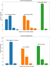
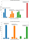
Introduction: Cryopreserved allograft heart valves (CAHV) show longer event-free survival compared to other types of protheses. However, all patients develop early and/or late allograft failure. Negative predictors are clinical, and there is a lack of evidence whether they correspond with the microscopic structure of CAHV. We assessed histopathological signs of structural degeneration, degree of cellular preservation, and presence of antigen-presenting cells (APC) in CAHV and correlated the changes with donor clinical characteristics, cryopreservation times, and CAHV types and diameters.
Methods: Fifty-seven CAHV (48 pulmonary, nine aortic) used for transplantation between November/2017 and May/2019 were included. Donor variables were age, gender, blood group, height, weight, and body surface area (BSA). Types and diameters of CAHV, cold ischemia time, period from decontamination to cryopreservation, and cryopreservation time were recorded. During surgery, arterial wall (n=56) and valvar cusp (n=20) samples were obtained from the CAHV and subjected to microscopy. Microscopic structure was assessed using basic staining methods and immunohistochemistry (IHC).
Results: Most of the samples showed signs of degeneration, usually of mild degree, and markedly reduced cellular preservation, more pronounced in aortic CAHV, correlating with arterial APC counts in both basic staining and IHC. There was also a correlation between the degree of degeneration of arterial samples and age, height, weight, and BSA of the donors. These findings were independent of preservation times.
Conclusion: CAHV show markedly reduced cellular preservation negatively correlating with the numbers of APC. More preserved CAHV may be therefore prone to stronger immune rejection.
Keywords: Allografts; Antigen Presenting Cells; Cryopreservation; Degeneration; Heart Valve; Histopathology; Tissue Donors.
Conflict of interest statement
No conflict of interest.
Figures


References
-
- Corno FA. Valved Conduits right ventricle to pulmonary artery for complex congenital heart defects. In: Cagini L, editor. Current Concepts in General Thoracic Surgery. 5. London: IntechOpen; 2012. - DOI
-
- Albert JD, Bishop DA, Fullerton DA, Campbell DN, Clarke DR. Conduit reconstruction of the right ventricular outflow tract. Lessons learned in a twelve-year experience. J Thorac Cardiovasc Surg. 1993;106(2):228–35. discussion 235-6. - PubMed
-
- Tweddell JS, Pelech AN, Frommelt PC, Mussatto KA, Wyman JD, Fedderly RT, et al. Factors affecting longevity of homograft valves used in right ventricular outflow tract reconstruction for congenital heart disease. Circulation. 2000;102(19 Suppl 3):III130–5. - PubMed
MeSH terms
LinkOut - more resources
Full Text Sources
Medical
