Human brain activity and functional connectivity as memories age from one hour to one month
- PMID: 35073239
- PMCID: PMC9308837
- DOI: 10.1080/17588928.2021.2021164
Human brain activity and functional connectivity as memories age from one hour to one month
Abstract
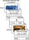
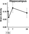
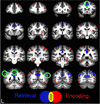
Theories of memory consolidation suggest the role of brain regions and connectivity between brain regions change as memories age. Human lesion studies indicate memories become hippocampus-independent over years, whereas animal studies suggest this process occurs across relatively short intervals, from days to weeks. Human neuroimaging studies suggest that changes in hippocampal and cortical activity and connectivity can be detected over these short intervals, but many of these studies examined only two time periods. We examined memory and fMRI activity for photos of indoor and outdoor scenes across four time periods to examine these neural changes more carefully. Participants (N = 21) studied scenes 1 hour, 1 day, 1 week, or 1 month before scanning. During scanning, participants viewed scenes, made old/new recognition memory judgments, and gave confidence ratings. Memory accuracy, confidence ratings, and response times changed with memory age. Brain activity in a widespread cortical network either increased or decreased with memory age, whereas hippocampal activity was not related to memory age. These findings were almost identical when effects of behavioral changes across time periods were minimized. Functional connectivity of the ventromedial prefrontal cortex with the posterior parietal cortex increased with memory age. By contrast, functional connectivity of the hippocampus with the parahippocampal cortex and fusiform gyrus decreased with memory age. In sum, we detected changes in cortical activity and changes in hippocampal and cortical connectivity with memory age across short intervals. These findings provide support for the predictions of systems consolidation and suggest that these changes begin soon after memories are formed.
Keywords: Consolidation; Functional Connectivity; functional magnetic resonance imaging; neuroimaging; retrograde memory.
Conflict of interest statement
The authors declare no conflict of interest.
Figures


Comment in
-
These things take time: what is the role of the hippocampus in recognition memory over extended delays?Cogn Neurosci. 2022 Jul;13(3-4):147-148. doi: 10.1080/17588928.2022.2076073. Epub 2022 May 16. Cogn Neurosci. 2022. PMID: 35575186
-
Challenges facing fMRI studies of systems consolidation.Cogn Neurosci. 2022 Jul;13(3-4):149-150. doi: 10.1080/17588928.2022.2076074. Epub 2022 May 16. Cogn Neurosci. 2022. PMID: 35575197
-
On the contribution of the ventromedial prefrontal cortex to the neural representation of past memories.Cogn Neurosci. 2022 Jul;13(3-4):154-155. doi: 10.1080/17588928.2022.2076072. Epub 2022 May 17. Cogn Neurosci. 2022. PMID: 35579493
-
Specifying 'where' and 'what' is critical for testing hippocampal contributions to memory retrieval.Cogn Neurosci. 2022 Jul;13(3-4):144-146. doi: 10.1080/17588928.2022.2076071. Epub 2022 May 18. Cogn Neurosci. 2022. PMID: 35586907 Free PMC article.
-
The devil may be in the details: The need for contextually rich stimuli in memory consolidation research.Cogn Neurosci. 2022 Jul;13(3-4):139-140. doi: 10.1080/17588928.2022.2076077. Epub 2022 May 19. Cogn Neurosci. 2022. PMID: 35587688
-
Evidence for the standard model, multiple trace theory, or the unified theory?Cogn Neurosci. 2022 Jul;13(3-4):151-153. doi: 10.1080/17588928.2022.2076663. Epub 2022 May 23. Cogn Neurosci. 2022. PMID: 35603813
-
Inquiring the librarian about the location of memory.Cogn Neurosci. 2022 Jul;13(3-4):134-136. doi: 10.1080/17588928.2022.2076075. Epub 2022 May 26. Cogn Neurosci. 2022. PMID: 35616221
-
Beyond the hippocampus: boundary conditions for cortical connectivity and activity over time.Cogn Neurosci. 2022 Jul;13(3-4):156-157. doi: 10.1080/17588928.2022.2080651. Epub 2022 May 27. Cogn Neurosci. 2022. PMID: 35621182
-
In search of systems consolidation.Cogn Neurosci. 2022 Jul;13(3-4):137-138. doi: 10.1080/17588928.2022.2080652. Epub 2022 Jun 4. Cogn Neurosci. 2022. PMID: 35659477
-
Changes in brain activity and connectivity as memories age.Cogn Neurosci. 2022 Jul;13(3-4):141-143. doi: 10.1080/17588928.2022.2076076. Epub 2022 Jun 13. Cogn Neurosci. 2022. PMID: 35695056
-
A way forward for design and analysis of neuroimaging studies of memory consolidation.Cogn Neurosci. 2022 Jul;13(3-4):158-164. doi: 10.1080/17588928.2022.2121274. Epub 2022 Sep 16. Cogn Neurosci. 2022. PMID: 36112016 Free PMC article.
References
-
- Bayley PJ, Gold JJ, Hopkins RO, & Squire LR (2005). The neuroanatomy of remote memory. Neuron, 46(5), 799–810. https://doi.org/S0896-6273(05)00394-6 [pii] 10.1016/j.neuron.2005.04.034 - DOI - PMC - PubMed
-
- Bontempi B, Laurent-Demir C, Destrade C, & Jaffard R (1999). Time-dependent reorganization of brain circuitry underlying long-term memory storage. Nature, 400, 671–675. - PubMed
Publication types
MeSH terms
Grants and funding
LinkOut - more resources
Full Text Sources
