. 2022 Feb 22;13(1):e0375221.
doi: 10.1128/mbio.03752-21. Epub 2022 Jan 25.

Bacterial Indole as a Multifunctional Regulator of Klebsiella oxytoca Complex Enterotoxicity

Affiliations

Bacterial Indole as a Multifunctional Regulator of Klebsiella oxytoca Complex Enterotoxicity

Nagender Ledala et al. mBio. .

Abstract

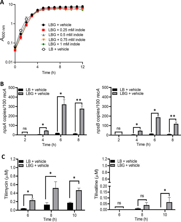
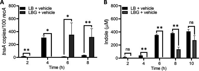
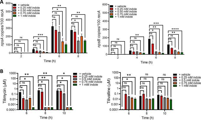
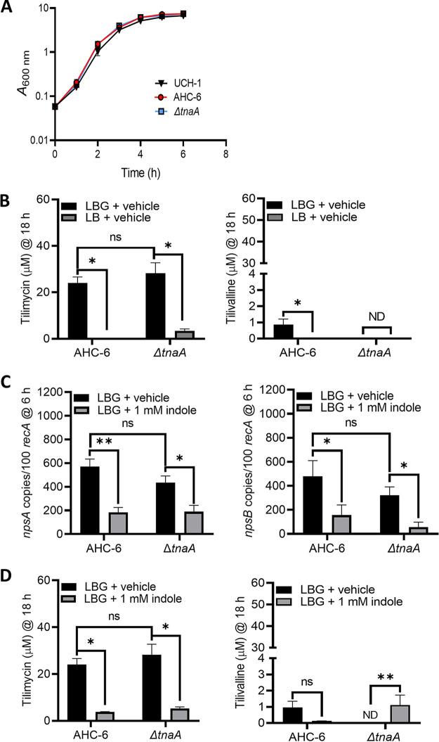
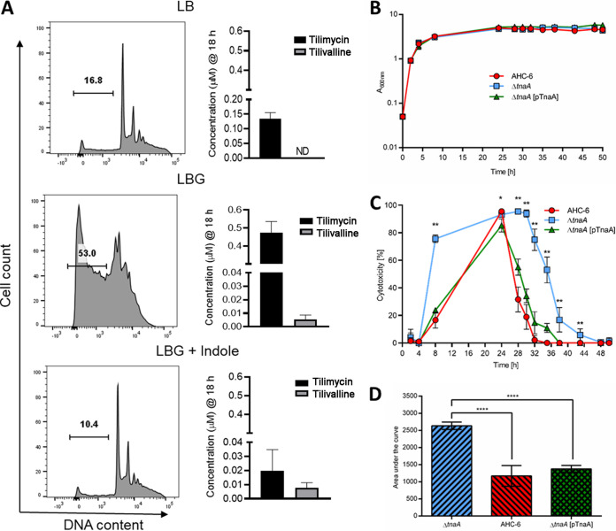
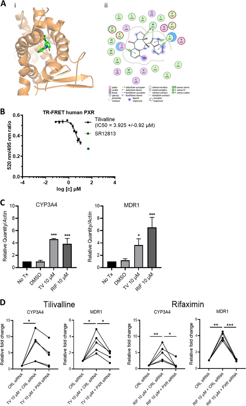
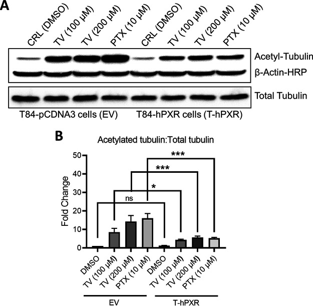
Gastrointestinal microbes respond to biochemical metabolites that coordinate their behaviors. Here, we demonstrate that bacterial indole functions as a multifactorial mitigator of Klebsiella grimontii and Klebsiella oxytoca pathogenicity. These closely related microbes produce the enterotoxins tilimycin and tilivalline; cytotoxin-producing strains are the causative agent of antibiotic-associated hemorrhagic colitis and have been associated with necrotizing enterocolitis of premature infants. We demonstrate that carbohydrates induce cytotoxin synthesis while concurrently repressing indole biosynthesis. Conversely, indole represses cytotoxin production. In both cases, the alterations stemmed from differential transcription of npsA and npsB, key genes involved in tilimycin biosynthesis. Indole also enhances conversion of tilimycin to tilivalline, an indole analog with reduced cytotoxicity. In this context, we established that tilivalline, but not tilimycin, is a strong agonist of pregnane X receptor (PXR), a master regulator of xenobiotic detoxification and intestinal inflammation. Tilivalline binding upregulated PXR-responsive detoxifying genes and inhibited tubulin-directed toxicity. Bacterial indole, therefore, acts in a multifunctional manner to mitigate cytotoxicity by Klebsiella spp.: suppression of toxin production, enhanced conversion of tilimycin to tilivalline, and activation of PXR. IMPORTANCE The human gut harbors a complex community of microbes, including several species and strains that could be commensals or pathogens depending on context. The specific environmental conditions under which a resident microbe changes its relationship with a host and adopts pathogenic behaviors, in many cases, remain poorly understood. Here, we describe a novel communication network involving the regulation of K. grimontii and K. oxytoca enterotoxicity. Bacterial indole was identified as a central modulator of these colitogenic microbes by suppressing bacterial toxin (tilimycin) synthesis and converting tilimycin to tilivalline while simultaneously activating a host receptor, PXR, as a means of mitigating tissue cytotoxicity. On the other hand, fermentable carbohydrates were found to inhibit indole biosynthesis and enhance toxin production. This integrated network involving microbial, host, and metabolic factors provides a contextual framework to better understand K. oxytoca complex pathogenicity.

Keywords: Klebsiella oxytoca complex; cytotoxin; indole; intestinal inflammation; pregnane X receptor.


Conflict of interest statement

The authors declare no conflict of interest.

Figures

FIG 1
FIG 1
Glucose induces cytotoxin synthesis by UCH-1. (A) UCH-1 was grown in LB broth alone or with glucose (LBG) containing indole at various concentrations with shaking at 37°C for 12 h (n = 3 to 8). (B) Effect of glucose on npsA and npsB expression. Transcript copy numbers of npsA and npsB (per 100 copies of recA transcript) in samples obtained at exponential (2 to 4 h) and postexponential (6 to 8 h) growth phases were determined by qRT-PCR (n = 3 to 8). (C) Tilimycin and tilivalline levels in postexponential growth phase culture supernatants determined by LC-MS (n = 3). The data are presented as mean values ± standard errors of the means. Statistical analysis was done by Mann-Whitney U test. Where stated, “vehicle” means addition of DMF (0.1%). *, P ≤ 0.05; **, P ≤ 0.01. ns, not significant.
FIG 2
FIG 2
Glucose represses expression of tnaA and indole biosynthesis by UCH-1. (A) tnaA transcript copy numbers (per 100 copies of recA transcripts) in samples obtained at exponential (2 to 4 h) and postexponential (6 to 8 h) growth phases determined by qRT-PCR (n = 3 to 8). (B) Indole levels in exponential- to postexponential-growth-phase culture supernatants determined by LC-MS (n = 3 to 7). The data are presented as mean values ± standard errors of the means. Statistical analysis was done by Mann-Whitney U test.*, P ≤ 0.05; **, P ≤ 0.01.
FIG 3
FIG 3
Exogenous indole represses cytotoxin synthesis by UCH-1. (A) Expression of the npsA and npsB genes in response to increasing concentrations of indole. Transcript copy numbers for npsA and npsB (per 100 copies of recA transcript) in samples obtained at exponential (2 to 4 h) and postexponential (6 to 8 h) growth phases were determined by qRT-PCR (n = 3 to 8). (B) Analysis of tilimycin and tilivalline metabolite levels in exponential- to postexponential-growth-phase culture supernatants by LC-MS (n = 3). The data are presented as the means values ± standard errors of the means. Statistical analysis was done by Kruskal-Wallis test followed by Dunn’s posttest. *, P ≤ 0.05; **, P ≤ 0.01; ***, P ≤ 0.001; ****, P ≤ 0.0001.
FIG 4
FIG 4
Glucose and indole reciprocally regulate cytotoxin synthesis by K. oxytoca strain AHC-6 and the AHC-6ΔtnaA mutant. (A) Comparison of UCH-1 growth with AHC-6 and AHC-6ΔtnaA in LB medium with glucose (LBG) at 37°C for 6 h (n = 3 to 7). (B) Analysis of tilimycin and tilivalline metabolite levels from 18-h culture supernatants of AHC-6 and AHC-6 ΔtnaA grown in LB medium with or without added glucose determined by LC-MS. ND, not detected (n = 3 to 8). (C) Effect of indole (1 mM) on npsA and npsB expression in AHC-6 and AHC-6 ΔtnaA grown in LBG. Transcript copy numbers for npsA and npsB (per 100 copies of recA transcript) in samples obtained at postexponential (6 h) growth phase (n = 6 or 7) were determined by RT-qPCR. (D) Effect of indole (1 mM) on tilimycin and tilivalline metabolite levels from 18-h culture supernatants of AHC-6 and AHC-6 ΔtnaA grown in LBG determined by LC-MS (n = 3 to 7). The data represent mean values ± standard errors of the means. Statistical analysis was done by Mann-Whitney U test. In samples where no tilivalline was detected, the detection limit by LC-MS was used for statistics. Where stated, “vehicle” means addition of DMF (0.1%). *, P ≤ 0.05; **, P ≤ 0.01.
FIG 5
FIG 5
Enterotoxicity of K. oxytoca complex is differentially regulated by glucose and indole. (A) Filtered supernatants from K. grimontii strain UCH-1 cultures, grown for 18 h in LB medium, LBG, or LBG plus 1.0 mM indole, were applied to T84 enterocytes. Shown are percentages of sub-G1 apoptotic cells following 72 h of exposure as determined by flow cytometry. Concentrations of tilimycin and tilivalline in the culture supernatants were determined by LC-MS (n = 3 to 8). The data are representative images or mean values ± standard errors of the means. (B) Growth comparison of indole-producing K. oxytoca AHC-6, the indole-deficient AHC-6 ΔtnaA, and complementing strain AHC-6 ΔtnaA (pTnaA) grown in CASO medium at 37°C (n = 3). The data represent mean values ± SD. (C) Cytotoxicity of filtered supernatants collected from cells used for panel B on HeLa cells (n = 3; measured in triplicates). Shown are reciprocal values (means ± SD) of surviving HeLa cells after treatment with 1/27 dilutions of supernatants. Statistical comparison (Mann-Whitney) of AHC-6 and AHC-6 ΔtnaA was done for each time point. *, P < 0.05; **, P < 0.01. (D) Statistical analysis (ANOVA, Tukey) of cytotoxicity over time. Compared are mean values (±SD) of calculated area under the curve. ****, P = 0.0001.
FIG 6
FIG 6
Indole represses tilimycin synthesis in vivo. C57BL/6NRj mice were infected with K. oxytoca AHC-6 (n = 4) or the indole-deficient ΔtnaA strain (n = 6). (A) K. oxytoca colonization levels were determined by plating fecal samples on CASO-kanamycin agar. Shown are CFU per gram of feces over time for individual mice colonized with AHC-6 or the ΔtnaA strain; black horizontal bars represent the geometric means. The inset shows a simplified comparison of geometric means for both strains over time in the same scale. (B) Fecal tilimycin and indole values were determined by LC-MS daily for each mouse. Plotted are mean ± SEM tilimycin and indole values for each group (AHC-6; ΔtnaA) and day. Dashed black lines represent the limit of quantification for indole (LOQ) and simultaneously indicate lack of quantifiable indole values for the ΔtnaA group. (C) Statistical significance of differences in total tilimycin amounts (area under the curve) generated by AHC-6 and the ΔtnaA mutant before (days 0 to 2) and after (days 2 to 6) indole production was determined by Mann-Whitney U test. *, P = 0.0333.
FIG 7
FIG 7
Tilivalline is a PXR agonist. (A) Docked complex of tilivalline with PXR. (i) Three-dimensional representation with PXR represented as ribbons and colored orange, while tilivalline is represented as licorice sticks and colored atom type (C, green; N, blue; and O, red). Hydrogen atoms have been removed for clarity. (ii) Schematic representation of the interactions of tilivalline with the ligand binding domain of PXR drawn using the ligand interaction module of MOE. The schematic legend at the bottom explains the types of interactions. (B) LanthaScreen TR-FRET PXR competitive binding assay. TR-FRET ratios (520/495 nm) are plotted against concentrations of tilivalline and SR12813. Half-maximal inhibitory concentrations (IC50s) were obtained from interpolated standard curves (sigmoidal, 4PL, variable slope); error bars show standard deviations (n = 3 or 4). (C) Relative mRNA levels for cyp3A4 and mdr1 in LS180 cells treated with tilivalline (TV) (10 μM), rifaximin (RIF) (10 μM; positive control), or DMSO (0.1% [vol/vol]; vehicle) determined by RT-qPCR. Data represent mean values ± standard errors of the means (n = 3 or 4). Tx, treatment. (D) Changes in cyp3A4 and mdr1 mRNA levels following treatment with TV (10 μM) or RIF (10 μM) in LS180 cells transfected with control (CRL) or PXR siRNA (n = 5). For panels C and D, statistical analysis was done by one-way ANOVA with multiple comparisons. *, P ≤ 0.05; **, P ≤ 0.01; ***, P ≤ 0.001.
FIG 8
FIG 8
PXR abrogates tilivalline-induced enterocyte tubulin acetylation. (A) Immunoblot of acetylated tubulin, total tubulin, and β-actin in T84 cells stably transfected with empty vector pCDNA3 (EV) or human PXR (hPXR). Cells were treated with tilivalline (100 μM and 200 μM) or paclitaxel (PTX) (10 μM), an established tubulin-acetylating/polymerizing agent whose toxic effects are modulated by PXR; cell lysates were transferred to nitrocellulose membranes and probed with antibodies specific for anti-acetylated tubulin, total tubulin, and β-actin. (B) Values of band intensities in EV and T-hPXR T84 cells with or without various treatments. The data represent the mean values ± standard errors of the means (n = 3). Statistical analysis was done by one-way ANOVA with multiple comparisons. *, P ≤ 0.05; ***, P ≤ 0.001.

References

    1. Kamada N, Chen GY, Inohara N, Nunez G. 2013. Control of pathogens and pathobionts by the gut microbiota. Nat Immunol 14:685–690. doi:10.1038/ni.2608. - DOI - PMC - PubMed
    1. Cameron EA, Sperandio V. 2015. Frenemies: signaling and nutritional integration in pathogen-microbiota-host interactions. Cell Host Microbe 18:275–284. doi:10.1016/j.chom.2015.08.007. - DOI - PMC - PubMed
    1. Jochum L, Stecher B. 2020. Label or concept—what is a pathobiont? Trends Microbiol 28:789–792. doi:10.1016/j.tim.2020.04.011. - DOI - PubMed
    1. Casadevall A. 2017. The pathogenic potential of a microbe. mSphere 2:e00015-17. doi:10.1128/mSphere.00015-17. - DOI - PMC - PubMed
    1. Kumar A, Sperandio V. 2019. Indole signaling at the host-microbiota-pathogen interface. mBio 10:e01031-19. doi:10.1128/mBio.01031-19. - DOI - PMC - PubMed

Publication types

MeSH terms

LinkOut - more resources