. 2022 Feb 22;13(1):e0366221.
doi: 10.1128/mbio.03662-21. Epub 2022 Jan 25.

N7-Methylation of the Coronavirus RNA Cap Is Required for Maximal Virulence by Preventing Innate Immune Recognition

Affiliations

N7-Methylation of the Coronavirus RNA Cap Is Required for Maximal Virulence by Preventing Innate Immune Recognition

Ruangang Pan et al. mBio. .

Abstract

The ongoing coronavirus (CoV) disease 2019 (COVID-19) pandemic caused by infection with severe acute respiratory syndrome CoV 2 (SARS-CoV-2) is associated with substantial morbidity and mortality. Understanding the immunological and pathological processes of coronavirus diseases is crucial for the rational design of effective vaccines and therapies for COVID-19. Previous studies showed that 2'-O-methylation of the viral RNA cap structure is required to prevent the recognition of viral RNAs by intracellular innate sensors. Here, we demonstrate that the guanine N7-methylation of the 5' cap mediated by coronavirus nonstructural protein 14 (nsp14) contributes to viral evasion of the type I interferon (IFN-I)-mediated immune response and pathogenesis in mice. A Y414A substitution in nsp14 of the coronavirus mouse hepatitis virus (MHV) significantly decreased N7-methyltransferase activity and reduced guanine N7-methylation of the 5' cap in vitro. Infection of myeloid cells with recombinant MHV harboring the nsp14-Y414A mutation (rMHVnsp14-Y414A) resulted in upregulated expression of IFN-I and ISG15 mainly via MDA5 signaling and in reduced viral replication compared to that of wild-type rMHV. rMHVnsp14-Y414A replicated to lower titers in livers and brains and exhibited an attenuated phenotype in mice. This attenuated phenotype was IFN-I dependent because the virulence of the rMHVnsp14-Y414A mutant was restored in Ifnar-/- mice. We further found that the comparable mutation (Y420A) in SARS-CoV-2 nsp14 (rSARS-CoV-2nsp14-Y420A) also significantly decreased N7-methyltransferase activity in vitro, and the mutant virus was attenuated in K18-human ACE2 transgenic mice. Moreover, infection with rSARS-CoV-2nsp14-Y420A conferred complete protection against subsequent and otherwise lethal SARS-CoV-2 infection in mice, indicating the vaccine potential of this mutant. IMPORTANCE Coronaviruses (CoVs), including SARS-CoV-2, the cause of COVID-19, use several strategies to evade the host innate immune responses. While the cap structure of RNA, including CoV RNA, is important for translation, previous studies indicate that the cap also contributes to viral evasion from the host immune response. In this study, we demonstrate that the N7-methylated cap structure of CoV RNA is pivotal for virus immunoevasion. Using recombinant MHV and SARS-CoV-2 encoding an inactive N7-methyltransferase, we demonstrate that these mutant viruses are highly attenuated in vivo and that attenuation is apparent at very early times after infection. Virulence is restored in mice lacking interferon signaling. Further, we show that infection with virus defective in N7-methylation protects mice from lethal SARS-CoV-2, suggesting that the N7-methylase might be a useful target in drug and vaccine development.

Keywords: N7-methylation; RNA cap structure; RNA methylation; SARS-CoV-2; coronavirus; immune response; interferons; nonstructural protein 14 (nsp14); type I interferon.


Conflict of interest statement

The authors declare no conflict of interest.

Figures
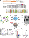
FIG 1
FIG 1
Sequence alignment of CoV-nsp14 and assessment of N7-MTase activity. (a) MHV-A59 genome organization and sequence alignment of the N7-MTase domain of nsp14 from several CoVs. The complete protein sequence of several CoV nsp14s was obtained from GenBank for representative α, β, and γ CoVs. The putative exonuclease (N terminus) and N7-methyltransferase (N7-MTase) (C terminus) domains are shown. Conserved residues within the N7-MTase are highlighted in red and blue, and their numbering corresponds to the location within MHV nsp14. MHV, mouse hepatitis virus strain A59; SARS-CoV, severe acute respiratory syndrome CoV; MERS-CoV, Middle East respiratory syndrome CoV; SARS-CoV-2, severe acute respiratory syndrome CoV 2; HCoV-229E, human coronavirus 229E; FIPV, feline infectious peritonitis virus; IBV, infectious bronchitis virus. The GenBank accession number is shown for each sequence. (b) Structure of MHV nsp14 with the substrates GpppA (in magenta) and SAM (in yellow) and residues (D330, N380, Y414, and N416) as indicated. MHV nsp14 is modeled based on SARS-CoV nsp14 (PDB accession no. 5C8S). Cyan, carbon; red, oxygen; blue, nitrogen. (c) Yeast complementation assay to examine the RNA cap guanosine-N7-methylation function of MHV and SARS-CoV-2 nsp14 mutants, as indicated. YBS40 was complemented by WT MHV nsp14 and the positive control, pYX232-HCM1, but not by MHV nsp14 mutants (with the mutation Y414A or Y414H, with D330A as a negative control). (d) TLC analysis of P1-resistant cap structures released from G*pppA-RNA (substrate) treated with bacterium-derived MHV nsp14-D330A, MHV nsp14-Y414A, and WT MHV nsp14. The positions of origin and migrations of m7G*pppA and G*pppA are indicated on the left (* indicates 32P labeled). (e) Infection of murine 17Cl-1 cells with rMHV or rMHVnsp14-Y414A at an MOI of 1 (left) or 0.001 (right). The titer of the virus was determined at the indicated time points. (f) GpppA-RNA was used to test the methylation activities of SARS-CoV-2 nsp14 and its mutants, as indicated. ctr, control. (g) In vitro N7-methylation assay using bacterially expressed MHV nsp14 or MHV nsp10 and MHV nsp16. Total poly(A)-containing RNAs were isolated from the culture supernatants of neuro-2A cells infected with rMHV and rMHVnsp14-Y414A. Incorporation of 3H was measured after treatment with the indicated proteins in the presence of 3H-labeled SAM. ns, not significant; ***, P < 0.001 (unpaired Student t test). Data are representative of three independent experiments (mean values ± SD).
FIG 2
FIG 2
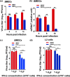
rMHVnsp14-Y414A-induced IFN-β expression and IFN signaling in primary cells. (a) Levels of IFN-β in L2 cell supernatants obtained at 12 hpi. (b) Levels of IFN-β in supernatants of infected bone marrow-derived dendritic cells (BMDCs) at the indicated time points. (c, d) RNA isolated from BMDCs infected with rMHV or rMHVnsp14-Y414A at 8 hpi and subjected to microarray analysis. (c) The heatmap shows 257 genes differentially expressed (DE) between rMHVnsp14-Y414A-infected BMDCs and mock- or rMHV-infected BMDCs. (d) Differentially expressed immune response genes. (e) Two micrograms of viral RNA were isolated from purified virions harvested from the supernatant and was transfected into BMDCs. Cells were analyzed for Ifn-β mRNA at 8, 12, and 16 hpi. (f) Fifty nanomolar RNA fragments (51 nt) from the 5′-UTR of the MHV-A59 genome with the indicated 5′-end sequences were transfected into BMDCs and analyzed for Ifn-β mRNA by qRT-PCR at 20 h posttransfection. Poly(I·C) was a positive control. ns, not significant; **, P < 0.01; ***, P < 0.001 (unpaired Student t test). Data are representative of three independent experiments (mean values ± SD). Rel., relative.
FIG 3
FIG 3
Diminished replication of rMHVnsp14-Y414A in BMDCs and increased IFN-β sensitivity. (a, b) Infection of BMDCs with rMHV or rMHVnsp14-Y414A at an MOI of 0.1. (a, b) Virus titers at the indicated time points were assessed by plaque assay (a) and by qRT-PCR with primers useful for detecting MHV mRNA7 (nucleocapsid gene) (b). Levels of mRNA7 relative to that of β-actin mRNA are expressed as 2–ΔCT[ΔCT = CT(mRNA7) − CT(β-actin)], where CT is the threshold cycle. (c, d) BMDCs (c) and L2 cells (d) were treated with the indicated dosages of IFN-β for 4 h before and after infection with rMHV or rMHVnsp14-Y414A (MOI of 1). Virus titers were measured 8 h after infection. ns, not significant; *, P < 0.05; **, P < 0.01; ***, P < 0.001 (unpaired Student t test). Data are representative of three independent experiments (mean values ± SD).
FIG 4
FIG 4
IFN-β expression in rMHVnsp14-Y414A-infected primary cells is dependent upon RLR activation. (a) BMDCs transfected with siRNA targeting RIG-I (left) or MDA5 (right) for 24 h, prior to infection with rMHV or rMHVnsp14-Y414A at an MOI of 0.1. Virus titers were measured by plaque assay at 16 hpi. (b, c) Infection of C57BL/6 (b) or MDA5-deficient (mda5−/−) (c) peritoneal macrophages with rMHV, rMHVnsp14-Y414A, or Sendai virus (SeV) as a positive control at an MOI of 1. The level of IFN-β in the cell supernatant was measured by ELISA at 12 hpi. (d, e) Infection of peritoneal macrophages derived from C57BL/6 (d) or mda5−/− (e) mice with rMHV or rMHVnsp14-Y414A at an MOI of 0.0001. Virus titers were measured by plaque assay. ns, not significant; *, P < 0.05; **, P < 0.01; ***, P < 0.001 (unpaired Student t test). Data are representative of three independent experiments (mean values ± SD).
FIG 5
FIG 5
rMHVnsp14-Y414A exhibits reduced pathogenicity in mice. Four-week-old C57/BL6 mice (n = 5) were inoculated intrahepatically (i.h.) (a, c, e) or intracranially (i.c.) (b, d, f) with 104 PFU of rMHV or rMHVnsp14-Y414A. (a, b) Mortality was monitored daily. (c, d) Levels of serum ALT were measured at 24, 72, and 120 hpi. (e, f) Livers of rMHV- or rMHVnsp14-Y414A- infected mice were stained with hematoxylin-eosin and examined for pathological changes at 1 and 3 days p.i. Arrowheads indicate inflammatory foci and tissue damage in livers of the infected mice. The inset shows higher magnification. Data are representative of two independent experiments (mean values ± SD). IU, international units.
FIG 6
FIG 6
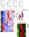
Early expression of IFN-β in sera and transcriptomic analysis in livers of rMHV- and rMHVnsp14Y414A-infected C57BL/6 mice. For IFN-β expression, IFN-β in the sera of 4-week-old C57BL/6 mice (six mice per each group) infected with rMHV or rMHVnsp14-Y414A was assayed at the indicated time points by ELISA after i.h. (a) or i.c. (b) inoculation. Data are representative of two independent experiments (mean values ± SD). For transcriptome analysis, 4-week-old C57BL/6 mice (3 mice per each group) were intrahepatically inoculated with rMHV and rMHVnsp14Y414A (7.5 × 105 PFU of virus per mouse). After 16 h and 24 h of infection, liver tissues of mice were collected for transcriptome sequencing. (c) A heatmap depicting the expression pattern of genes significantly upregulated and downregulated in each group is shown. (d) Numbers of upregulated and downregulated genes differentially expressed between infected mice and naive mice. Criteria used for differential expression analysis are a q value of <0.05 and a |log2 fold change| of >1. (e) Differentially expressed immune response-related genes.
FIG 7
FIG 7
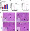
Restriction of rMHVnsp14-Y414A replication is restored in Ifnar−/− BMDCs and mice. (a) Infection of Ifnar−/− BMDCs with rMHV or rMHVnsp14-Y414A at an MOI of 0.1. Virus titers at the indicated time points were assessed by plaque assay. (b, c) Four-week-old Ifnar−/− mice were infected i.h. (b) or i.c. (c) and monitored for survival. (d) Histopathological analysis of liver tissues of Ifnar−/− mice infected with rMHV or rMHVnsp14-Y414A at 1 dpi. Arrowheads indicate large perivascular infiltrates detected at 1 dpi in rMHV- or rMHVnsp14-Y414A-infected mice. (b to d) Data are representative of 10 mice per group. ns, not significant; *, P < 0.05 (unpaired Student t test). Data are representative of two independent experiments (mean values ± SD).
FIG 8
FIG 8
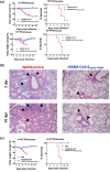
rSARS-CoV-2nsp14-Y414A is attenuated and protects mice against lethal SARS-CoV-2 challenge. (a) Eight-week-old K18-hACE2 transgenic mice were intranasally (i.n.) infected with 103 PFU (upper) or 104 PFU (bottom) of rSARS-CoV-2 or rSARS-CoV-2nsp14-Y420A, respectively (n = 5). Body weights (left) and mortality (right) were monitored daily. (b) Lungs of mice infected with rSARS-CoV-2 or rSARS-CoV-2nsp14-Y420A were stained with hematoxylin-eosin and examined for pathological changes at 3 and 10 days p.i. Black arrows indicated inflammatory cell infiltrates. (c) Eight-week-old K18-hACE2 transgenic mice were inoculated with 1 × 104 PFU of rSARS-CoV-2nsp-Y420A or phosphate-buffered saline (PBS) (n = 5). At 40 dpi, animals were i.n. challenged with a lethal dose of 5 × 104 PFU of rSARS-CoV-2. Weight change (left) and survival (right) were observed daily. Means with SD are shown (n = 5).

References

    1. Braciale TJ, Hahn YS. 2013. Immunity to viruses. Immunol Rev 255:5–12. doi:10.1111/imr.12109. - DOI - PMC - PubMed
    1. Totura AL, Baric RS. 2012. SARS coronavirus pathogenesis: host innate immune responses and viral antagonism of interferon. Curr Opin Virol 2:264–275. doi:10.1016/j.coviro.2012.04.004. - DOI - PMC - PubMed
    1. Furuichi Y, Shatkin AJ. 2000. Viral and cellular mRNA capping: past and prospects. Adv Virus Res 55:135–184. doi:10.1016/s0065-3527(00)55003-9. - DOI - PMC - PubMed
    1. Cowling VH, Cole MD. 2010. Myc regulation of mRNA cap methylation. Genes Cancer 1:576–579. doi:10.1177/1947601910378025. - DOI - PMC - PubMed
    1. Daffis S, Szretter KJ, Schriewer J, Li J, Youn S, Errett J, Lin TY, Schneller S, Zust R, Dong H, Thiel V, Sen GC, Fensterl V, Klimstra WB, Pierson TC, Buller RM, Gale M, Jr, Shi PY, Diamond MS. 2010. 2'-O methylation of the viral mRNA cap evades host restriction by IFIT family members. Nature 468:452–456. doi:10.1038/nature09489. - DOI - PMC - PubMed

Publication types