. 2022 May;10(5):1356-1364.e2.
doi: 10.1016/j.jaip.2021.12.038. Epub 2022 Jan 21.

Effective Anti-SARS-CoV-2 Immune Response in Patients With Clonal Mast Cell Disorders

Affiliations

Effective Anti-SARS-CoV-2 Immune Response in Patients With Clonal Mast Cell Disorders

Julien Rossignol et al. J Allergy Clin Immunol Pract. 2022 May.

Abstract

Background: Mast cells are key players in innate immunity and the TH2 adaptive immune response. The latter counterbalances the TH1 response, which is critical for antiviral immunity. Clonal mast cell activation disorders (cMCADs, such as mastocytosis and clonal mast cell activation syndrome) are characterized by abnormal mast cell accumulation and/or activation. No data on the antiviral immune response in patients with MCADs have been published.

Objective: To study a comprehensive range of outcomes in patients with cMCAD with PCR- or serologically confirmed coronavirus disease 2019 and to characterize the specific anti-severe acute respiratory syndrome coronavirus 2 (SARS-CoV-2) immune response in this setting.

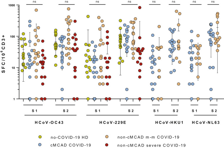
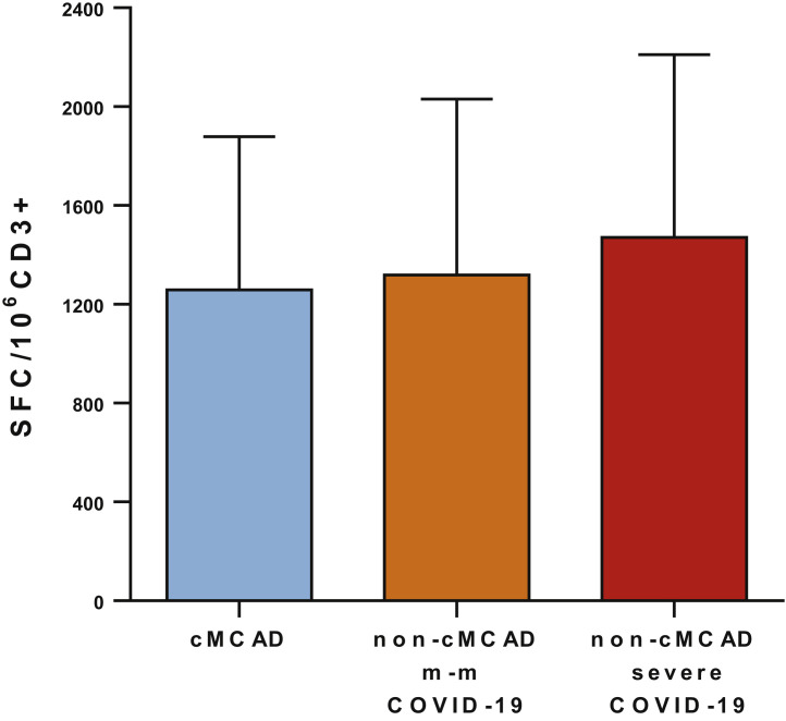
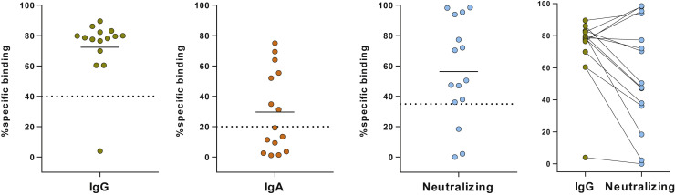
Methods: Clinical follow-up and outcome data were collected prospectively over a 12-month period by members of the French Centre de Référence des Mastocytoses rare disease network. Anti-SARS-CoV-2-specific T-cell activity was measured with an ELISA, and humoral responses were evaluated by assaying circulating levels of specific IgG, IgA, and neutralizing antibodies.

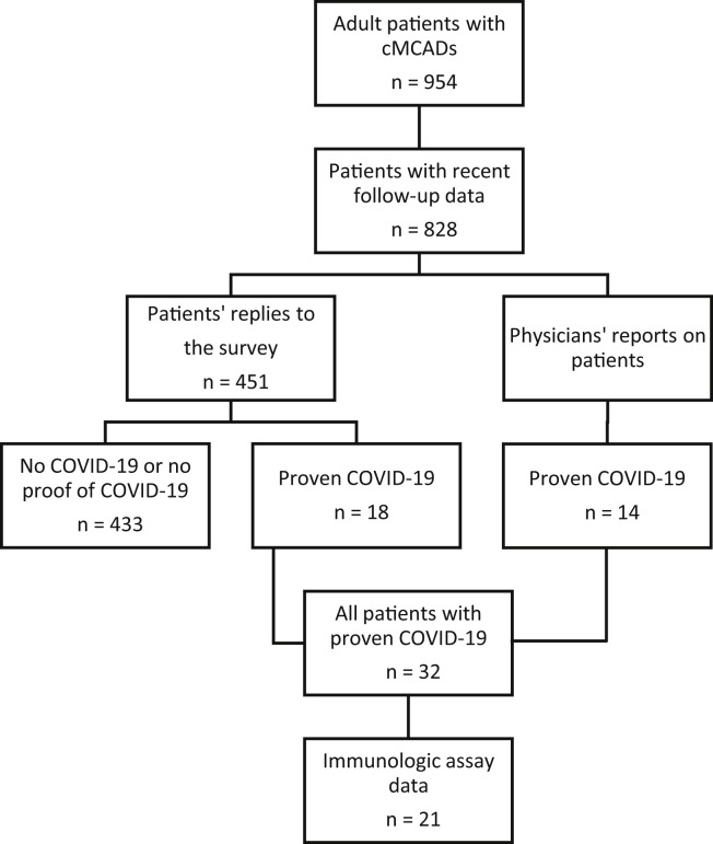
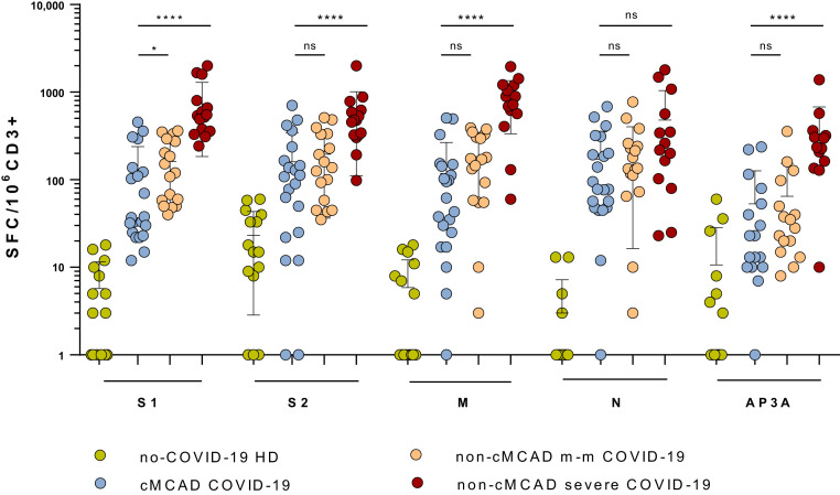
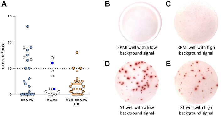
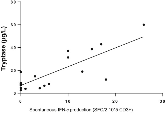
Results: Overall, 32 patients with cMCAD were evaluated. None required noninvasive or mechanical ventilation. Two patients were admitted to hospital for oxygen and steroid therapy. The SARS-CoV-2-specific immune response was characterized in 21 of the 32 patients. Most had high counts of circulating SARS-CoV-2-specific, IFN-γ-producing T cells and high titers of neutralizing antispike IgGs. The patients frequently showed spontaneous T-cell IFN-γ production in the absence of stimulation; this production was correlated with basal circulating tryptase levels (a marker of the mast cell burden).

Conclusions: Patients with cMCADs might not be at risk of severe coronavirus disease 2019, perhaps due to their spontaneous production of IFN-γ.

Keywords: B cells; COVID-19; Clonal mast cell activation syndrome; Mast cell activation disorders; Mast cells; Mastocytosis; SARS-CoV-2; T cells.


Figures

Figure 1
Figure 1
Flowchart for the selection of patients with cMCADs and proven COVID-19.
Figure 2
Figure 2
Quantification of specific anti–SARS-CoV-2 T-cell responses, using an ELISpot assay. The tested SARS-CoV-2 peptide pools were derived from a peptide scan through the SARS-CoV-2 spike glycoprotein (S1: N-terminal fragment, S2: C-terminal fragment), membrane protein (M), nucleoprotein (N), and ORF3a protein (AP3a). The negative controls were PBMCs in culture medium alone, and the positive controls were PHA and the CEFX Ultra SuperStim Pool. Results were expressed as the number of SFCs/106 CD3+ T cells after subtraction of the background values from wells with nonstimulated cells. All differences between the non–COVID-19 and COVID-19 groups were statistically significant (P < .001). cMCADs, Convalescent patients with cMACDs; HD, healthy donors; non-cMCAD m-m COVID-19, control patients without cMACD convalescing from a mild to moderate form of COVID-19 (n = 17); non-cMCAD non-COVID-19, non-cMCAD, non-COVID-19 controls (n = 15); non-cMCADs severe COVID-19, control patients without cMACD convalescing from a severe form of COVID-19 (n = 15). ns, nonsignificant. ∗P < .05; ∗∗∗∗P < .0001.
Figure 3
Figure 3
Spontaneous IFN-γ production by PBMCs from patients, as measured in an ELISpot assay. (A) Graphic representation of the ELISpot assays. cMCADs, patients with cMCADs; non-cMCAD control groups: MCAS, patients with idiopathic mast cell activation syndrome; CTR: non-cMCAD/non-MCAS control patients convalescing from COVID-19 (n = 32); HD, healthy donors. Empty circles: no history of COVID-19. Filled circles: history of COVID-19. Pictures of ELISpot assays. (B) A well with nonstimulated PBMCs from a COVID-19 non-cMCAD control. (C) A well with nonstimulated PBMCs from a patient with COVID-19 and cMCAD. (D) A well with PBMCs from a COVID-19 non-cMCAD control after stimulation for 18 to 20 hours with individual 15-mer 11-aa overlapping peptide pools derived from the SARS-CoV-2 N-terminal fragment spike protein. (E) A well with PBMCs from a patient with COVID-19 and cMCAD after stimulation with individual 15-mer 11-aa overlapping peptide pools derived from the SARS-CoV-2 N-terminal fragment spike protein.
Figure 4
Figure 4
Correlation between basal tryptase level (μg/L) and spontaneous IFN-γ production (SFCs/2 × 105 CD3), as observed in ELISpot assays. n = 24 patients with CM, MIS, and ISM. Linear regression: r2 = 0.44 (P < .0004). CM, Cutaneous mastocytosis; MIS, mastocytosis in the skin.
Figure E1
Figure E1
T-cell responses to common coronaviruses in patients with cMCAD (n = 17), and patients without cMCAD with a history of mild to moderate COVID-19 (n = 17) or severe COVID-19 (n = 15), or healthy donors (n = 15). Identification of HCoV-OC43–, HCoV-229E–, HCoV-HKU1–, and HCoV-NL63–specific T-cell responses, using ELISpot assays. Results were expressed as SFCs/106 CD3+ T cells after subtraction of background values from wells containing nonstimulated cells. cMCADs, Patients with cMCADs, convalescing from mild to moderate (m-m) or severe COVID-19; ns, nonsignificant
Figure E2
Figure E2
T-cell responses to the CEFX Ultra SuperStim Pool. cMCADs, Patients with MCADs, convalescing from mild to moderate (m-m) or severe COVID-19.
Figure E3
Figure E3
Two left-most panels: serologic status for anti–SARS-CoV-2 IgA and IgG antibodies, determined in an S-flow assay. The dashed line indicates the positivity threshold. Third panel: Percentage IgG neutralizing ability, determined in a viral pseudoparticle assay. Fourth panel: relationship between the IgG titer and the neutralizing activity.

References

    1. Valent P., Akin C., Metcalfe D.D. Mastocytosis: 2016 updated WHO classification and novel emerging treatment concepts. Blood. 2017;129:1420–1428. - PMC - PubMed
    1. Lim K., Tefferi A., Lasho T.L., Finke C., Patnaik M., Butterfield J.H., et al. Systemic mastocytosis in 342 consecutive adults: survival studies and prognostic factors. Blood. 2009;113:5727–5736. - PubMed
    1. Gotlib J., Pardanani A., Akin C., Reiter A., George T., Hermine O., et al. International Working Group-Myeloproliferative Neoplasms Research and Treatment (IWG-MRT) & European Competence Network on Mastocytosis (ECNM) consensus response criteria in advanced systemic mastocytosis. Blood. 2013;121:2393–2401. - PMC - PubMed
    1. Helmy Y.A., Fawzy M., Elaswad A., Sobieh A., Kenney S.P., Shehata A.A. The COVID-19 pandemic: a comprehensive review of taxonomy, genetics, epidemiology, diagnosis, treatment, and control. J Clin Med. 2020;9:1225. - PMC - PubMed
    1. Hadjadj J., Yatim N., Barnabei L., Corneau A., Boussier J., Smith N., et al. Impaired type I interferon activity and inflammatory responses in severe COVID-19 patients. Science. 2020;369:718–724. - PMC - PubMed

Publication types

Substances