Review
. 2022 May 27;12(5):a041176.
doi: 10.1101/cshperspect.a041176.

Lymphedema and Obesity

Affiliations
Review

Lymphedema and Obesity

Christopher L Sudduth et al. Cold Spring Harb Perspect Med. .

Abstract

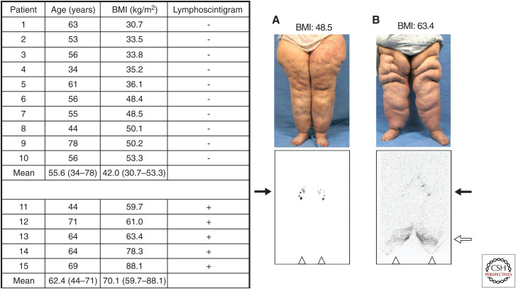
Lymphedema results from inadequate lymphatic function. Extreme obesity can cause lower extremity lymphedema, termed "obesity-induced lymphedema (OIL)." OIL is a form of secondary lymphedema that may occur once an individual's body mass index (BMI) exceeds 40. The risk of lymphatic dysfunction increases with elevated BMI and is almost universal once BMI exceeds 60. Obesity has a negative impact on lymphatic density in subcutaneous tissue, lymphatic endothelial cell proliferation, lymphatic leakiness, collecting-vessel pumping capacity, and clearance of macromolecules. Lymphatic fluid unable to be taken up by lymphatic vessels results in increased subcutaneous adipose deposition, fibrosis, and worsening obesity. Individuals with OIL are in an unfavorable cycle of weight gain and lymphatic injury. The fundamental treatment for OIL is weight loss.


Figures

Figure 1.
Figure 1.
Original 2012 cohort of obese patients with bilateral lower extremity enlargement and a referral diagnosis of “lymphedema.” All patients with a body mass index (BMI) ≤ 53.3 had a negative lymphoscintigram, indicating normal lymphatic function of the lower limbs. Each patient with a BMI ≥ 59.7 had a positive study (i.e., delayed transport of radiolabeled tracer and/or dermal backflow), showing abnormal lymphatic drainage consistent with lymphedema. (A) 55-yr-old woman (patient #7) with normal transit of technetium-99m (Tc-99m) filtered sulfur colloid to the inguinal nodes 45 min following intradermal injection into the feet. (B) 64-yr-old woman (patient #13) with delayed tracer uptake into the inguinal nodes and dermal backflow 5 h after injection of the radiolabeled colloid. Inguinal nodes (black arrows); dermal backflow in distal lower extremities (white arrow); injection sites in feet (triangles). (Figure from Greene et al. 2012; reprinted, with permission, from the Massachusetts Medical Society © 2012.)
Figure 2.
Figure 2.
Follow-up 2015 cohort of obese patients with or without lymphedema. Patients without a history of massive weight loss and a body mass index (BMI) <50 kg/m2 had normal lower extremity lymphatic function. Individuals with a BMI >60 kg/m2 had abnormal findings on lymphoscintigraphy consistent with lymphedema. A BMI threshold between 50 kg/m2 and 60 kg/m2 appeared to exist, at which point lymphatic dysfunction occurs. Patients on the left with a normal lymphoscintigram result exhibited inguinal lymph node uptake of technetium Tc-99m sulfur colloid 45 min after injection into the feet. Subjects on the right with an abnormal lymphoscintigram result showed delayed transport of tracer and/or dermal backflow on 3-h images. Black arrows identify inguinal lymph nodes, and the white arrow marks dermal backflow. (Figure from Greene et al. 2015; reprinted, with permission, from Wolters Kluwer Health © 2015.)
Figure 3.
Figure 3.
Patients with obesity-induced lymphedema (OIL) are at risk for developing areas of massive localized lymphedema (MLL). (A) Patient with a body mass index (BMI) of 70 has OIL and MLL of the left thigh. (B) The abnormal lymphoscintigram exhibits reduced inguinal lymph node uptake of technetium-99m sulfur colloid and dermal backflow in the left leg. (C) The area of MLL is significantly improved after the BMI was lowered to 30 following a surgical-weight loss procedure. (Figure from Maclellan et al. 2017; reprinted, with permission, from Elsevier © 2017.)

References

    1. Arngrim N, Simonsen L, Holst JJ, Bülow J. 2013. Reduced adipose tissue lymphatic drainage of macromolecules in obese subjects: a possible link between obesity and local tissue inflammation. Int J Obes 37: 748–750. 10.1038/ijo.2012.98 - DOI - PubMed
    1. Blum KS, Karaman S, Proulx ST, Ochsenbein AM, Luciani P, Leroux JC, Wolfrum C, Detmar M. 2014. Chronic high-fat diet impairs collecting lymphatic vessel function in mice. PLoS ONE 9: e94713. 10.1371/journal.pone.0094713 - DOI - PMC - PubMed
    1. Brorson H, Ohlin K, Olsson G, Nilsson M. 2006. Adipose tissue dominates chronic arm lymphedema following breast cancer: an analysis using volume rendered CT images. Lymphat Res Biol 4: 199–210. 10.1089/lrb.2006.4404 - DOI - PubMed
    1. Farshid G, Weiss SW. 1998. Massive localized lymphedema in the morbidly obese: a histologically distinct reactive lesion simulating liposarcoma. Am J Surg Pathol 22: 1277–1283. 10.1097/00000478-199810000-00013 - DOI - PubMed
    1. García Nores GD, Cuzzone DA, Albano NJ, Hespe GE, Kataru RP, Torrisi JS, Gardenier JC, Savetsky IL, Aschen SZ, Nitti MD, et al. 2016. Obesity but not high-fat diet impairs lymphatic function. Int J Obes (Lond) 40: 1582–1590. 10.1038/ijo.2016.96 - DOI - PMC - PubMed