

The use of 3D printed models for the pre-operative planning of surgical correction of pediatric hip deformities: a case series and concise review of the literature
- PMID: 35075078
- PMCID: PMC8823571
- DOI: 10.23750/abm.v92i6.11703
The use of 3D printed models for the pre-operative planning of surgical correction of pediatric hip deformities: a case series and concise review of the literature
Abstract
Background and aim: Three-dimensional (3D) printing is prevailing in surgical planning of complex cases. The aim of this study is to describe the use of 3D printed models during the surgical planning for the treatment of four pediatric hip deformity cases. Moreover, pediatric pelvic deformities analyzed by 3D printed models have been object of a concise review.
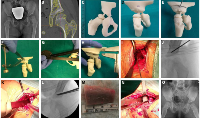
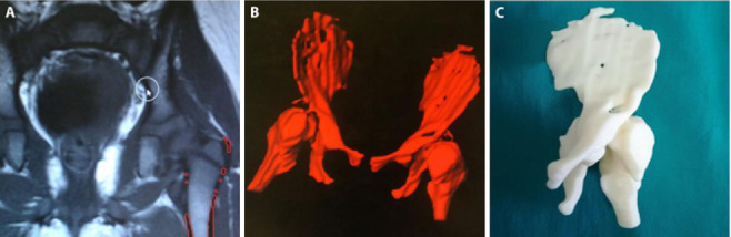
Methods: All treated patients were females, with an average age of 5 years old. Patients' dysplastic pelvises were 3D-printed in real scale using processed files from Computed Tomography (CT) or Magnetic Resonance Imaging (MRI). Data about 3D printing, surgery time, blood loss and fluoroscopy have been recorded.
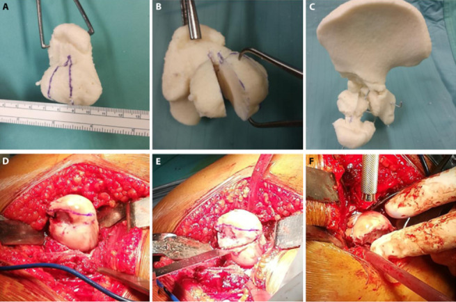
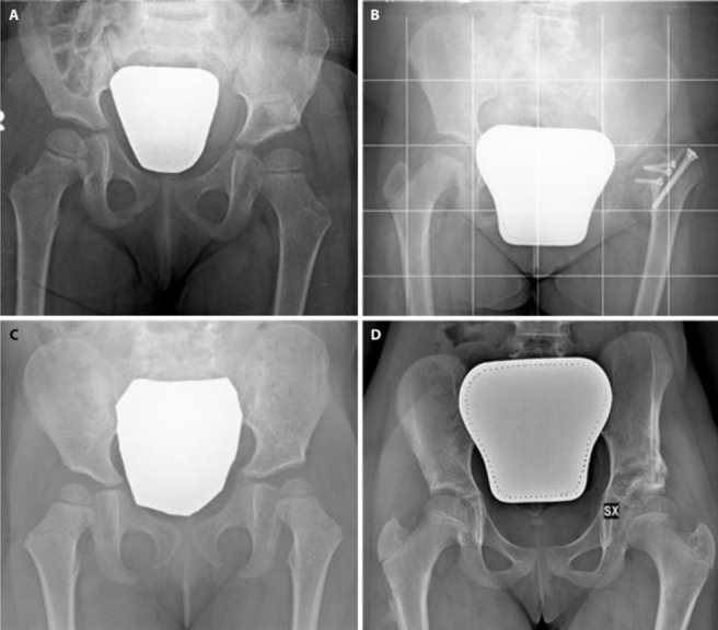
Results: The Zanoli-Pemberton or Ganz-Paley osteotomies were performed on the four 3D printed models, then the real surgery was performed in the operating room. Time and costs to produce 3D printed models were respectively on average 17:26 h and 34.66 €. The surgical duration took about 87.5 min while the blood loss average was 1.9 ml/dl. Fluoroscopy time was 21 sec. MRI model resulted inaccurate and more difficult to produce. 10 papers have been selected for the concise literature review.
Conclusions: 3D printed models have proved themselves useful in the reduction of surgery time, blood loss and ionizing radiation, as well as they have improved surgical outcomes. 3D printed model is a valid tool to deepen the complex anatomy and orientate surgical choices by allowing surgeons to carefully plan the surgery.
Conflict of interest statement
Each author declares that he or she has no commercial associations (e.g. consultancies, stock ownership, equity interest, patent/licensing arrangement etc.) that might pose a conflict of interest in connection with the submitted article.
Figures
References
-
- Caffrey JP, Jeffords ME, Farnsworth CL, Bomar JD, Upasani VV. Comparison of 3 Pediatric Pelvic Osteotomies for Acetabular Dysplasia Using Patient-specific 3D-printed Models. J Pediatr Orthop. 2019;39(3):e159–e164. - PubMed
-
- Murphy RF, Kim YJ. Surgical Management of Pediatric Developmental Dysplasia of the Hip. J Am Acad Orthop Surg. 2016;24(9):615–624. - PubMed
-
- Coppa V, Marinelli M, Specchia N. Unilateral uniplanar modular external fixator for percutaneous proximal femoral osteotomy in children: surgical technique. Eur J Orthop Surg Traumatol. 2019;29(1):205–211. - PubMed
-
- Paley D, Feldman DS. Femoral Head Reduction Osteotomy. In: Saran N, Hamdy R.C, editors. Pediatric Pelvic and Proximal Femoral Osteotomies: a case-based approach. Springer; 2018. pp. 379–420.
Publication types
MeSH terms
LinkOut - more resources
Full Text Sources
