

Primary extra-skeletal Ewing's sarcoma presenting as an epidural Soft Tissue Lesion causing cauda equina syndrome in an adolescent girl: a case report
- PMID: 35075107
- PMCID: PMC8786895
- DOI: 10.1038/s41394-021-00474-7
Primary extra-skeletal Ewing's sarcoma presenting as an epidural Soft Tissue Lesion causing cauda equina syndrome in an adolescent girl: a case report
Abstract
Introduction: Primary epidural Ewing's sarcoma in the lumbar spinal canal is a rare condition and very few cases are reported in the literature.
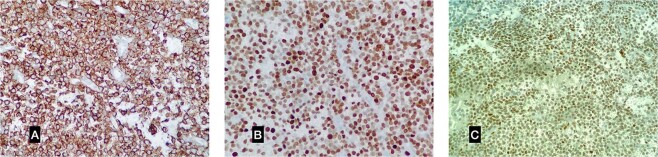
Case report: A fifteen-year-old girl presented with low backache associated with sudden onset of weakness and radiculopathy of both lower limbs for 10 days, bowel and bladder involvement for 3 days. Physical examination revealed grade 0/5 power and absent sensations below L4 dermatomal level and perianal region (ASIA A). Plantar reflex was mute bilaterally. Magnetic resonance imaging revealed an extradural lesion within the spinal canal at the L3-L4 level. The patient underwent an emergency posterior decompression, extradural lesion excision and instrumented stabilization L3-L5. Based on histopathological examination of the tissue specimen, we diagnosed the lesion as Ewing sarcoma.
Discussion: Primary extra-skeletal Ewing's sarcoma presenting as an epidural lesion in the lumbar spine is a rare clinical entity that should be considered as a differential for spinal epidural lesions. Treatment for such cases is almost always an early surgical intervention due to its rapid onset and compressive neurological symptoms. Wide decompression with instrumented fusion and excision of the lesion followed by chemo and radiotherapy are recommended.
© 2022. The Author(s), under exclusive licence to International Spinal Cord Society.
Conflict of interest statement
The authors declare no competing interests.
Figures


References
-
- Ewing J. Diffuse Endothelioma of Bone. Proc. New York Path. Soc. 1921;21:17–24.
-
- Esiashvili N, Goodman M, Marcus RB. Changes in incidence and survival of Ewing sarcoma patients over the past 3 decades: Surveillance Epidemiology and End Results data. J Pediatr Hematol/Oncol. 2008;30:425–30. - PubMed
-
- Ries L, Percy C, Bunin G. Introduction—SEER pediatric monograph. Cancer Incid survival Child adolescents: US SEER Program. 1975;1995:1–15.
-
- Tefft M, Vawter G, Mitus A. Paravertebral “round cell” tumors in children. Radiology. 1969;92:1501–9. - PubMed
-
- Raney RB, Asmar L, Newton WA, Bagwell C, Breneman JC, Crist W, et al. Ewing’s sarcoma of soft tissues in childhood: a report from the Intergroup Rhabdomyosarcoma Study, 1972 to 1991. J Clin Oncol. 1997;15:574–82.. - PubMed
Publication types
MeSH terms
LinkOut - more resources
Full Text Sources
