. 2022 Jan 24;13(1):75.
doi: 10.1038/s41419-022-04519-z.

Loss of NEDD4 causes complete XY gonadal sex reversal in mice

Affiliations

Loss of NEDD4 causes complete XY gonadal sex reversal in mice

Simon P Windley et al. Cell Death Dis. .

Abstract

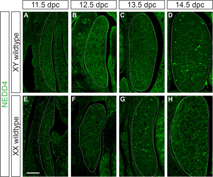
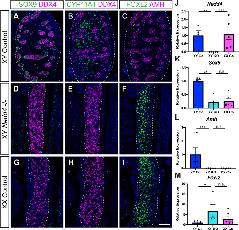
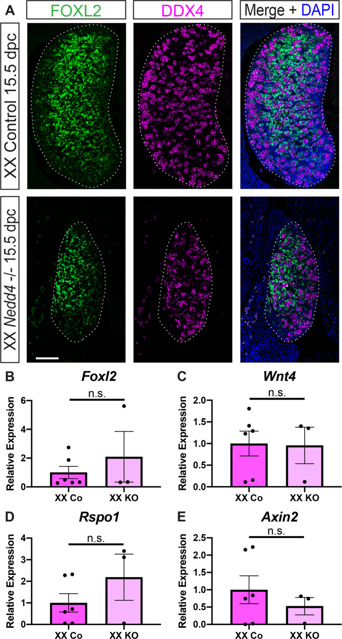
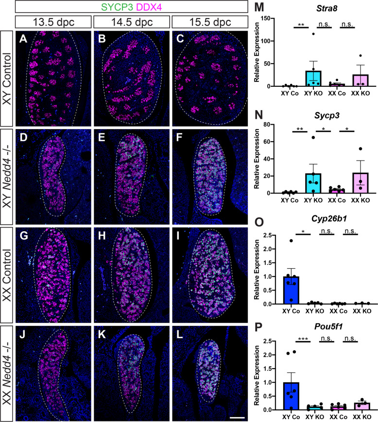
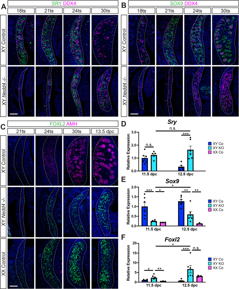
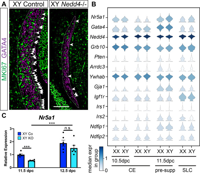
Gonadogenesis is the process wherein two morphologically distinct organs, the testis and the ovary, arise from a common precursor. In mammals, maleness is driven by the expression of Sry. SRY subsequently upregulates the related family member Sox9 which is responsible for initiating testis differentiation while repressing factors critical to ovarian development such as FOXL2 and β-catenin. Here, we report a hitherto uncharacterised role for the ubiquitin-protein ligase NEDD4 in this process. XY Nedd4-deficient mice exhibit complete male-to-female gonadal sex reversal shown by the ectopic upregulation of Foxl2 expression at the time of gonadal sex determination as well as insufficient upregulation of Sox9. This sex reversal extends to germ cells with ectopic expression of SYCP3 in XY Nedd4-/- germ cells and significantly higher Sycp3 transcripts in XY and XX Nedd4-deficient mice when compared to both XY and XX controls. Further, Nedd4-/- mice exhibit reduced gonadal precursor cell formation and gonadal size as a result of reduced proliferation within the developing gonad as well as reduced Nr5a1 expression. Together, these results establish an essential role for NEDD4 in XY gonadal sex determination and development and suggest a potential role for NEDD4 in orchestrating these cell fate decisions through the suppression of the female pathway to ensure proper testis differentiation.


Conflict of interest statement

The authors declare no competing interests.

Figures

Fig. 1
Fig. 1. NEDD4 is ubiquitously expressed in the developing XY gonad.
Section immunofluorescence on XY (AD) and XX (EH) wildtype embryos from 11.5 dpc to 14.5 dpc stained for NEDD4 (green). Gonads are denoted by a white dotted line. The anterior pole of each gonad is positioned at the top of each panel. Scale bars = 100 μm.
Fig. 2
Fig. 2. Complete male-to-female sex reversal in XY Nedd4-/- mice.
AI Section immunofluorescence on 14.5 dpc XY Nedd4-/- embryos alongside XY and XX littermate controls stained for Sertoli cell markers SOX9 (green in A, D, G) and AMH (magenta in C, F, I), Leydig cell marker CYP11A1 (green in B, E, H), germ cell marker DDX4 (magenta in A, B, D, E, G, H) and granulosa cell marker FOXL2 (green in C, F, I). Gonads are denoted by a white dotted line. The anterior pole of each gonad is positioned at the top of each panel. Scale bars = 100 μm. RT-qPCR analyses of Nedd4 (J), Sox9 (K), Amh (L) and Foxl2 (M) expression at 14.5 dpc on XY Nedd4-/- gonads (KO) (n = 5), XY controls (Co) (n = 6) and XX controls (n = 6). Values are expressed relative to the XY controls. Mean ± SEM; t-test, n.s. not significant, *p < 0.05, **p < 0.01, ***p < 0.001.
Fig. 3
Fig. 3. Normal ovarian somatic cell development in XX Nedd4-/- mice.
A Section immunofluorescence on 15.5 dpc XX Nedd4-/- embryos alongside XX littermate controls stained for ovarian somatic cells markers FOXL2 (green) and germ cell marker DDX4 (magenta). The anterior pole of each gonad is positioned at the top of each panel. Gonads are denoted by a white dotted line. Scale bars = 100 μm. (BE) RT-qPCR analyses of Foxl2 (B), Wnt4 (C), Rspo1 (D) and Axin2 (E) expression at 14.5 dpc on XX Nedd4-/- gonads (KO) (n = 3) and XX controls (n = 6). Values are expressed relative to the XX controls. Mean ± SEM; t-test, n.s. not significant.
Fig. 4
Fig. 4. Germ cells in XX Nedd4-/- mice enter meiosis.
AL Section immunofluorescence on 13.5 dpc, 14.5 dpc and 15.5 dpc XY and XX Nedd4-/- embryos alongside XY and XX littermate controls stained for germ cell marker DDX4 (magenta) and meiosis marker SYCP3 (green). Gonads are denoted by a white dotted line. The anterior pole of each gonad is positioned at the top of each panel. Scale bars = 100 μm. RT-qPCR analyses of Stra8 (M), Sycp3 (N), Cyp26b1 (O) and Pou5f1 (P) expression at 14.5 dpc on XY Nedd4-/- gonads (XY KO) (n = 5), XX KO gonads (n = 3), XY controls (Co) (n = 6) and XX controls (n = 6). Values are expressed relative to the XY controls. Mean ± SEM; t-test; n.s. not significant, *p < 0.05, **p < 0.01, ***p < 0.001.
Fig. 5
Fig. 5. Impaired Sertoli cell differentiation and ectopic FOXL2 in XY Nedd4-/- mice.
Section immunofluorescence for the male sex determinant SRY (green) (A) and Sertoli cell marker SOX9 (green) (B) co-labelled with the germ cell marker DDX4 (magenta) on XY Nedd4-/- embryos and XY wildtype controls between 18ts and 30ts. C Section immunofluorescence for the granulosa cell marker FOXL2 (green) and Sertoli cell marker AMH (magenta) on XY Nedd4-/- embryos alongside XY and XX controls between 21ts and 13.5 dpc. The anterior pole of each gonad is positioned at the top of each panel. Gonads are denoted by a white dotted line. Scale bars = 100μm. RT-qPCR analyses of Sry (D), Sox9 (E) and Foxl2 (F) expression at 11.5 dpc (17–19ts - gonads + mesonephros) and 12.5 dpc (gonads only) on XY Nedd4-/- (KO) (light blue; n = 6), XY controls (Co) (dark blue; n = 6) and XX controls (pink; n = 3). Values are expressed relative to 11.5 dpc XY controls. Mean ± SEM; t-test; n.s. not significant, *p < 0.05, **p < 0.01, ***p < 0.001.
Fig. 6
Fig. 6. Impaired proliferation and decreased Nr5a1 expression in XY Nedd4-/- mice.
A Section immunofluorescence on 11.5 dpc (18 tail somites [ts]) XY Nedd4-/- embryos and XY controls stained for gonadal somatic cell marker GATA4 (magenta) and proliferation marker MKI67 (green). The anterior pole of each gonad is positioned at the top of each panel. Gonads are denoted by a white dotted line. MKI67-positive cells within the coelomic epithelium are highlighted by white arrowheads. Scale bar = 100μm. B Violin plots showing the expression of gonadal progenitor markers Nr5a1 and Gata4 as well as Nedd4 and Nedd4 interacting proteins in mouse gonadal cells expressing both Nr5a1 and Gata4 at E10.5 and E11.5. XX and XY expression levels are shown separately. Expression levels are shown in log-normalised counts. Colour indicates median expression in the group according to the scale shown in the left bottom corner. CE coelomic epithelium, pre-supp pre-supporting cells, SLC supporting-like cells. C RT-qPCR analyses of Nr5a1 at 11.5 dpc (17–19ts, gonads + mesonephroi) and 12.5 dpc (30ts, gonads only) on XY Nedd4-/- gonads (KO) (light blue; n = 6) and XY controls (Co) (dark blue; n = 6). Values are expressed relative to 11.5 dpc XY controls. Mean ± SEM; t-test; n.s. not significant, ***p < 0.001.

References

    1. Hu YC, Okumura LM, Page DC. Gata4 is required for formation of the genital ridge in mice. PLoS Genet. 2013;9:e1003629. - PMC - PubMed
    1. Bullejos M, Koopman P. Spatially dynamic expression of Sry in mouse genital ridges. Developmental Dyn. 2001;221:201–5. - PubMed
    1. Wilhelm D, Martinson F, Bradford S, Wilson MJ, Combes AN, Beverdam A, et al. Sertoli cell differentiation is induced both cell-autonomously and through prostaglandin signaling during mammalian sex determination. Dev Biol. 2005;287:111–24. - PubMed
    1. Sekido R, Lovell-Badge R. Sex determination involves synergistic action of SRY and SF1 on a specific Sox9 enhancer. Nature. 2008;453:930–4. - PubMed
    1. Chaboissier MC, Kobayashi A, Vidal VI, Lutzkendorf S, van de Kant HJ, Wegner M, et al. Functional analysis of Sox8 and Sox9 during sex determination in the mouse. Development. 2004;131:1891–901. - PubMed

Publication types

Substances