. 2022 Jan 24;5(1):89.
doi: 10.1038/s42003-021-02998-y.

Planar polarity in primate cone photoreceptors: a potential role in Stiles Crawford effect phototropism

Affiliations

Planar polarity in primate cone photoreceptors: a potential role in Stiles Crawford effect phototropism

Anna Verschueren et al. Commun Biol. .

Abstract

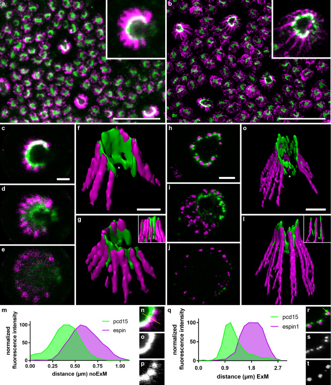
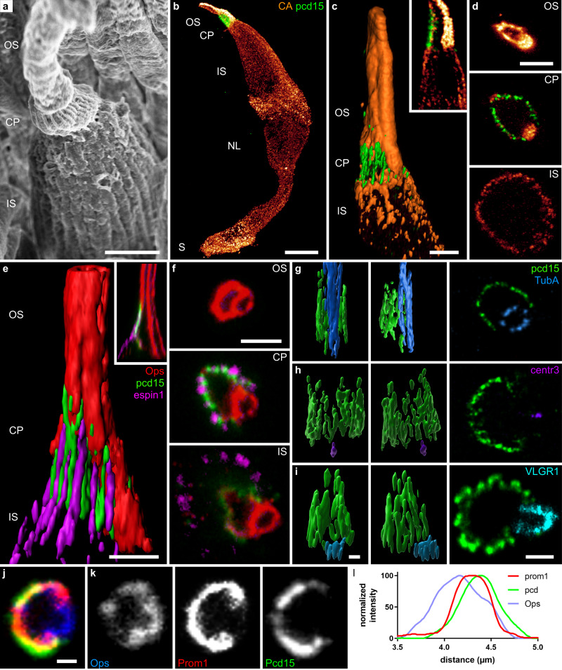
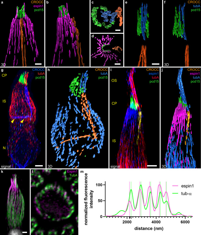
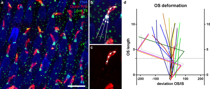
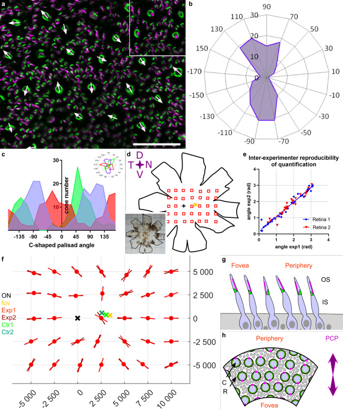
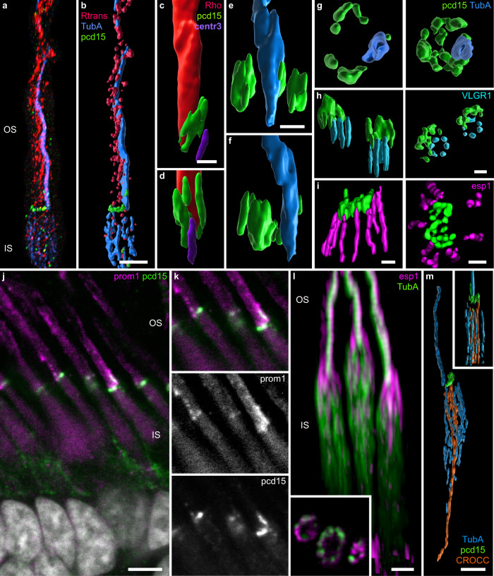
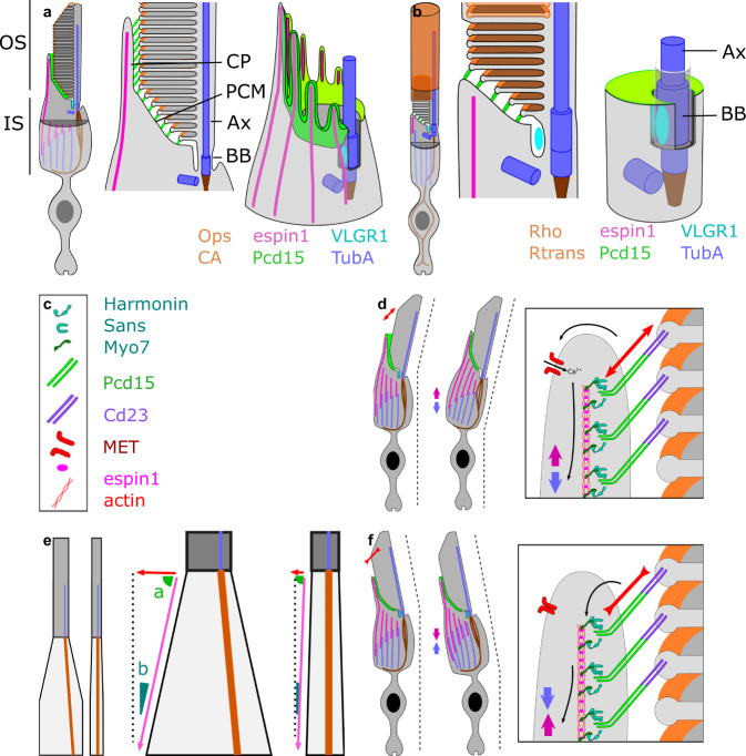
Human cone phototropism is a key mechanism underlying the Stiles-Crawford effect, a psychophysiological phenomenon according to which photoreceptor outer/inner segments are aligned along with the direction of incoming light. However, such photomechanical movements of photoreceptors remain elusive in mammals. We first show here that primate cone photoreceptors have a planar polarity organized radially around the optical center of the eye. This planar polarity, based on the structure of the cilium and calyceal processes, is highly reminiscent of the planar polarity of the hair cells and their kinocilium and stereocilia. Secondly, we observe under super-high resolution expansion microscopy the cytoskeleton and Usher proteins architecture in the photoreceptors, which appears to establish a mechanical continuity between the outer and inner segments. Taken together, these results suggest a comprehensive cellular mechanism consistent with an active phototropism of cones toward the optical center of the eye, and thus with the Stiles-Crawford effect.


Conflict of interest statement

The authors declare no competing interests.

Figures

Fig. 1
Fig. 1. High magnification of cone calyceal processes by expansion microscopy.
Photoreceptor mosaics were examined on flat mounts of the retina by classic confocal microscopy (a, cg, mp) and by expansion microscopy (b, hl, qt). Photoreceptors were immunolabeled for espin 1 (magenta) and prodocadherin 15 (pcd15, green). al Immunolabeling of cone photoreceptors visualized in the photoreceptor mosaic as z-projections (a, b), on confocal optical sections from the outer to the inner segment (ce, hj) or on 3D reconstructions from different viewpoints (fg, kl). mt Comparison of fluorescence profiles along a single calyceal process, showing the greater resolution of expansion microscopy (rt) than of classic confocal microscopy (np) for espin 1 and Pcd15 immunostaining (numerical data in supplementary data 1). The profile section (yellow line) is shown on the isolated calyceal processes (n, r). Scale bars represent 10 µm in (a) and (b), and 1 µm in (c) to (l) (after division by the 4.5 expansion factor in b, hl).
Fig. 2
Fig. 2. Calyceal C-shaped palisade around the cone cilium.
a Scanning electron microscopy of a primate cone showing the outer and inner segment as well as the calyceal process around the base of the outer segment. 3D reconstructions (b, c, e gi), reconstructed coronal slices (c and e inserts) and optical sections (d, f, last image of gi, and jk) of cone photoreceptors immunolabeled for the R/G cone opsin (red in a, dg; blue in l), espin 1 (magenta e, f), Protocadherin15 (green), cone arrestin (orange in bd), prominin 1 (red in j), acetylated alpha tubulin (blue in g), centrin 3 (purple in h), and VLGR1 (cyan in i). l normalized intensity across the cellular border. The Pcd15 staining is located in a position external to prominin 1. Numerical data in supplementary data 2. OS outer segment, CP calyceal processes, IS inner segment, NL nuclear layer, S synapse, arrowhead: pcd15 staining expression around the periciliary membrane at the base of the cilia. Scale bars represent 2 µm in (a, c, d) and 1 µm in (ej), and 10 µm in (b).
Fig. 3
Fig. 3. Cone cytoskeleton and inner/outer segment alignment.
3D reconstructions (ak, reconstructed volumes in af, h, and j), and an optical slice (l) of cones immunolabeled for espin 1 (magenta in all reconstructed volumes: a, b, d, j; blue in fluorescence 3D image I and magenta in fluorescence 3D image k), Protocadherin 15 (pcd15, green), acetylated tubulin alpha (tuba, blue in all reconstructed volumes: c, e, f, h, j; red in fluorescence 3D images in g and i, green in k and l) rootletin (CROCC, orange in all reconstructed volumes: c, e, f, h, and j, blue in fluorescence 3D image g and yellow in fluorescence 3D image i). a, b, d and c, e, f represent three perpendicular reconstitutions of the proteins. g yellow arrowheads underline the rootletin anchor. An optical slice in the mid-inner segment is shown for a single cone in l, the alternation between espin 1 and tubulin is readily visible both visually and on the fluorescence profile (m, numerical data in supplementary data 3). OS outer segment, CP calyceal processes, IS inner segment, N nucleus. Scale bars represent 0.5 µm in (c), 1 µm in (a, d, e), and 2 µm in (g, I, and k).
Fig. 4
Fig. 4. Cone inner/outer segment alignment.
ac Retinal flat-mount immunolabeled for the R/G cone opsin (red), Protocadherin 15 (pcd15, green) and peanut lectin agglutinin (PNA, blue). Definition of the angles between the inner and outer segments on an individual cone (b, c). d Graph showing the deviation of the angle from linearity for a population of 20 cone outer/inner segments, as a function of the distance to the base of immunostaining for Pcd15 (in degree). The dotted line represented the mean calyceal processes length. (numerical data in supplementary data 4). The scale bar in (a) represents 20 μm.
Fig. 5
Fig. 5. Planar polarity of cone photoreceptors in the macaque retina.
a Flat-mounted retina immunostained for protocadherin 15 (pcd15, green) and acetylated tubulin alpha (alpha-Tub, magenta), showing the directions vectors for all the cones in the image. Each cone vector is defined by the C-shaped symmetric axis of the Pcd15 staining and the direction toward the opening of the C-shape defined by the acetylated tubulin alpha staining, as illustrated in the inset. (b) Polar plot of the vectors on a retinal image (0.25 mm²) showing a clear alignment along a preferential planar axis. c Graphs representing the distributions of vector directions for 3 independent retinas, showing that all retinal samples had two peaks in opposite directions (numerical data in supplementary data 5). df Cone vector directions for the primate retina are shown in d. The planar axes are illustrated on the matrix (f) for all sample points located on the schematic representation of the retina (d). For each location, two experimenters independently placed individual cone vectors and calculated the mean planar axis. e Mean planar axis angle, demonstrating the reproducibility of the technique. (numerical data in supplementary data 6) f Graph providing the angles of the planar axis obtained for macaque 1 and for each observed retinal location in red (different hues for the datasets of different experimenters). Each planar axis was calculated from a minimum of 50 individual cone vector angles. The optic nerve, fovea, and both reconstructed centers (from the two different datasets) are represented by black, yellow, and green crosses, respectively. (macaque n2 in supplementary figure, and numerical data in supplementary data 7). g, h Schematic representation of the planar polarity organization discovered (g: view as a retinal radial section. Cones appear tilted toward the fovea. h: view as a flat-mounted retina. C cones, R rods, Green calyceal processes, magenta cilia). See Supplementary figure for comparison of macaque 1 and 2.
Fig. 6
Fig. 6. Junction of the outer and inner segments of rod photoreceptors.
3D reconstructions (ai, l, m) and optical sections (inset l) of rod photoreceptors immunolabeled for protocadherin-15 (pcd15, green in all images), acetylated alpha tubulin (TubA, blue in all reconstructed volumes: b, e, f, g, m, blue in fluorescent 3D image a, and green in fluorescent 3D image l), centrin 3 (violet in c, d), rhodopsin (red in c, d), transducin (red in a, b), espin 1 (magenta in I, l), VLGR1 (cyan in h), all examined by expansion microscopy (but l). j, k Double-labeling for prominin 1 and pcd15 on retinal slices (noExM). Scale bars after scaling down to take expansion into account are as follows: for a, b, l, m: 2 µm, c: 1 µm, di: 0.5 µm), j: 5 µm.
Fig. 7
Fig. 7. Schematic diagram of the distribution of USH proteins, other mechanical proteins, and cilium-associated proteins in rod and cone photoreceptors.
(a: cone, b rod). Schematic reconstruction of cone 3D inner/outer segment junction structure, showing: full photoreceptor view, a detailed transverse view, and a 3D reconstitution of the apical structure of the inner segment (outer segment remove for visual clarity). e Difference between cones and rods in inner-to-outer segment diameter ratio, leading to different effects of identical shortenings of the inner segment. d, f hypothesis for outer segment tilting leading to a change in inner segment alignment, c legend of the symbols are used in panels d and f. Ax axoneme, BB basal body, CP calyceal process, PCM periciliary membrane, OS outer segment, IS inner segment.

References

    1. Stiles WS. The luminous efficiency of monochromatic rays entering the eye pupil at different points and a new colour effect. Proc. R. Soc. Lond. Ser. B Biol. Sci. 1937;123:90–118.
    1. Laties, A. M. & Enoch, J. M. An analysis of retinal receptor orientation I. Angular relationship of neighboring photoreceptors. Investig. Ophtalmol. 10, 69–77 (1971). - PubMed
    1. Laties AM, Liebman PA, Campbell CE. Photoreceptor orientation in the primate eye. Nature. 1968;218:172–173. - PubMed
    1. Alan AM. Histological techniques for study of photoreceptor orientation. Tissue Cell. 1969;1:63–81. - PubMed
    1. Applegate MA, Bonds AB. Induced movement of receptor alignment toward a new pupillary aperture. Investig. Ophthalmol. Vis. Sci. 1981;21:869–873. - PubMed

Publication types

MeSH terms