

A U-Net Approach to Apical Lesion Segmentation on Panoramic Radiographs
- PMID: 35075428
- PMCID: PMC8783705
- DOI: 10.1155/2022/7035367
A U-Net Approach to Apical Lesion Segmentation on Panoramic Radiographs
Abstract
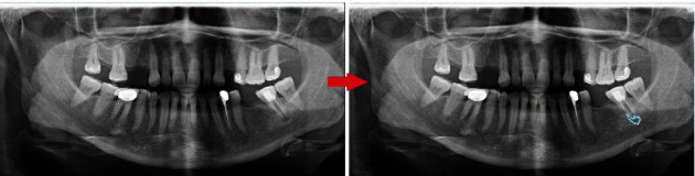
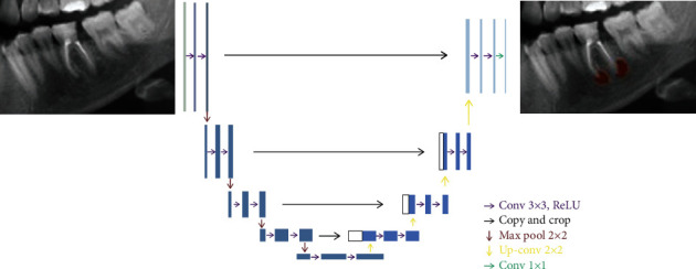
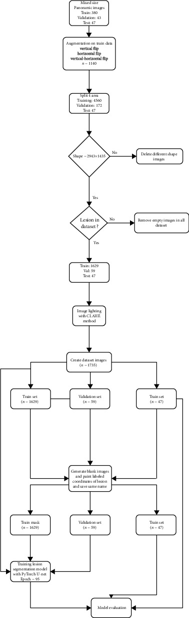
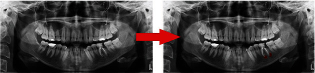
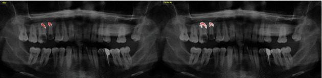
The purpose of the paper was the assessment of the success of an artificial intelligence (AI) algorithm formed on a deep-convolutional neural network (D-CNN) model for the segmentation of apical lesions on dental panoramic radiographs. A total of 470 anonymized panoramic radiographs were used to progress the D-CNN AI model based on the U-Net algorithm (CranioCatch, Eskisehir, Turkey) for the segmentation of apical lesions. The radiographs were obtained from the Radiology Archive of the Department of Oral and Maxillofacial Radiology of the Faculty of Dentistry of Eskisehir Osmangazi University. A U-Net implemented with PyTorch model (version 1.4.0) was used for the segmentation of apical lesions. In the test data set, the AI model segmented 63 periapical lesions on 47 panoramic radiographs. The sensitivity, precision, and F1-score for segmentation of periapical lesions at 70% IoU values were 0.92, 0.84, and 0.88, respectively. AI systems have the potential to overcome clinical problems. AI may facilitate the assessment of periapical pathology based on panoramic radiographs.
Copyright © 2022 Ibrahim S. Bayrakdar et al.
Conflict of interest statement
The authors declare that there is no conflict of interest regarding the publication of this paper.
Figures
References
-
- Ridao-Sacie C., Segura-Egea J., Fernández-Palacín A., Bullón-Fernández P., Ríos-Santos J. Radiological assessment of periapical status using the periapical index: comparison of periapical radiography and digital panoramic radiography. International Endodontic Journal . 2007;40(6):433–440. doi: 10.1111/j.1365-2591.2007.01233.x. - DOI - PubMed
-
- Patel S., Durack C. Essential Endodontology: Prevention and Treatment of Apical Periodontitis . Third Edition. Blackwell; 2019. Radiology of apical periodontitis. Essential endodontology: prevention and treatment of apical periodontitis; pp. 179–210.
MeSH terms
LinkOut - more resources
Full Text Sources
