

Post-COVID Wernicke's presenting as bilateral vision loss
- PMID: 35075437
- PMCID: PMC8769919
- DOI: 10.1016/j.ajoc.2022.101271
Post-COVID Wernicke's presenting as bilateral vision loss
Abstract
Purpose: This case report represents poor nutritional intake and vomiting secondary to COVID-19 resulting in Wernicke's syndrome and blindness.
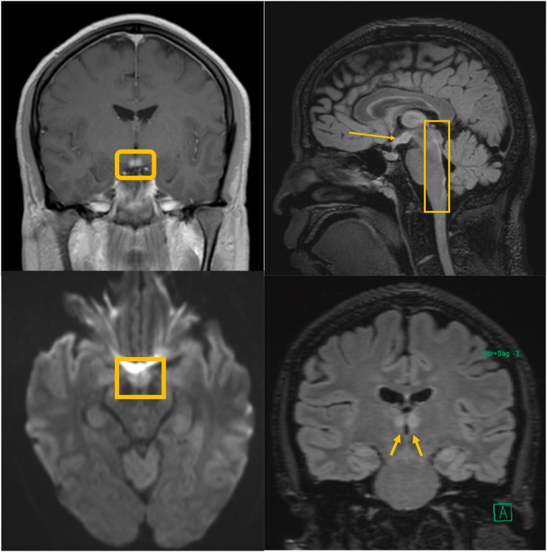
Observations: We report the case of a 36 year old with a post-COVID episode of acute-subacute onset bilateral blindness ultimately diagnosed as Wernicke's syndrome based on MRI findings and clinical response to high dose IV thiamine supplementation.
Conclusions and importance: Given this patient's dramatic presentation of no light perception vision in both eyes and resolution of symptoms with treatment, it is reasonable to consider thiamine deficiency in any individual who presents with acute-subacute onset vision loss, particularly when the history is suggestive of potential nutritional deficiency.
Keywords: COVID-19; Neuro-ophthalmology; Neurology; Nutrition; Optic neuropathy; SARS-CoV-2.
© 2022 Published by Elsevier Inc.
Conflict of interest statement
No authors has any conflicts of interest.
Figures
References
-
- Sinha S., Kataria A., Kolla B.P., Thusius N., Loukianova L.L. Wernicke encephalopathy—clinical pearls. Mayo Clin Proc. 2019;94(6):1065–1072. doi: 10.1016/j.mayocp.2019.02.018. https://search.datacite.org/works/10.1016/j.mayocp.2019.02.018 - DOI - DOI - PubMed
-
- De Wardener H.E., long mb, durh bl. Cerebral beriberi (wernicke's encephalopathy): review of 52 cases in a Singapore prisoner-of-war hospital. Lancet. 1947;249(6436):11–17. doi: 10.1016/S0140-6736(47)91272-5. http://www.sciencedirect.com/science/article/pii/S0140673647912725 - DOI - PubMed
Publication types
LinkOut - more resources
Full Text Sources
Miscellaneous
