. 2022 Feb 22;23(3):299-314.
doi: 10.1093/ehjci/jeab293.

Clinical applications of cardiac computed tomography: a consensus paper of the European Association of Cardiovascular Imaging-part I

Affiliations

Clinical applications of cardiac computed tomography: a consensus paper of the European Association of Cardiovascular Imaging-part I

Gianluca Pontone et al. Eur Heart J Cardiovasc Imaging. .

Erratum in

Abstract

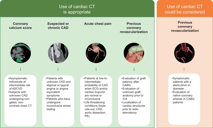
Cardiac computed tomography (CT) was introduced in the late 1990's. Since then, an increasing body of evidence on its clinical applications has rapidly emerged. From an initial emphasis on its technical efficiency and diagnostic accuracy, research around cardiac CT has now evolved towards outcomes-based studies that provide information on prognosis, safety, and cost. Thanks to the strong and compelling data generated by large, randomized control trials, the scientific societies have endorsed cardiac CT as pivotal diagnostic test for the management of appropriately selected patients with acute and chronic coronary syndrome. This consensus document endorsed by the European Association of Cardiovascular Imaging is divided into two parts and aims to provide a summary of the current evidence and to give updated indications on the appropriate use of cardiac CT in different clinical scenarios. This first part focuses on the most established applications of cardiac CT from primary prevention in asymptomatic patients, to the evaluation of patients with chronic coronary syndrome, acute chest pain, and previous coronary revascularization.

Keywords: acute chest pain; chronic coronary syndrome; coronary artery bypass graft; coronary calcium; coronary computed tomography angiography; coronary stent.


Figures

None
Clinical applications of cardiac CT. For more details, please see Table 1, which summarizes the main applications of cardiac CT. ASCVD, atherosclerotic cardiovascular disease; CABG, coronary artery by-pass graft; CAD, coronary artery disease; CT, computed tomography; ECG, electrocardiogram; ICA, invasive coronary angiography; PE, pulmonary embolism.
Figure 1
Figure 1
Coronary artery calcium assessment. (A) A non-enhanced, ECG-triggered axial CT scan was acquired in a 78-year-old man to measure the calcific plaque burden. (B) Tube potential was set to 120 kV resulting into a total DLP of 101.26 mGy*cm. (C and D) Image analysis was performed using a dedicated software, which automatically identified structures with a density ≥130 HU and highlighted them in green. Subsequently, coronary arteries were manually segmented (LM: turquoise, LAD: pink, LCX: yellow). (E and F) Total Agatston score (E) and per-vessel Agatston score (F) were calculated and correlated to age-matched cohorts to stratify patient’s risk. ECG, electrocardiogram; CT, computed tomography; DLP, dose length product; LAD, left anterior descending artery; LCX, left circumflex artery; LM, left main.
Figure 2
Figure 2
CCTA in symptomatic patients with suspected CAD. (A and B) A 56-year-old man underwent CCTA due to atypical chest pain after non-diagnostic exercise test. Single-phase, prospectively ECG-triggered axial CCTA was performed using a wide-detector CT scanner which allowed the coverage of the whole heart in a single beat (A), with a total DLP of 63.49 mGy*cm (B). (C–H) A non-calcific plaque (arrow) was detected in the proximal LAD as shown on curved MPR (C), straight MPR (D), cross-sectional views of the vessel (E and F) and volume rendering reconstruction (G and H). The yellow overlay in (F) indicates the non-calcific plaque and the associated severe stenosis (70–99%) resulting into a MLA of 0.5 mm2. CAD, coronary artery disease; CCTA, coronary computed tomography angiography; CT, computed tomography; DLP, dose length product; ECG, electrocardiogram; LAD, left anterior descending artery; MLA, minimal lumen area; MPR, multiplanar reconstruction.
Figure 3
Figure 3
CCTA in symptomatic patients with acute chest pain. (A–F) A 47-year-old woman with a history of hypertension was admitted to the emergency department for atypical chest pain. While physical exam and ECG were unremarkable (C), blood test results showed mild increase of hsTnI. No significant delta and/or ST-T changes were detected on serial assessments. CCTA showed no disease of LCX (A) and RCA (B) and demonstrated sub-occlusion of the mid LAD due to partially calcific plaque, as shown in the volume rendering reconstruction (D, arrow) and straight MPR (E, arrow). Dedicated plaque analysis software identified the fibroadipose (pink) and calcific (yellow) components of the plaque (F). (G and H) Invasive coronary angiography confirmed the sub-occlusion of mid LAD (G, arrow), which was treated with PCI+DES (H, arrow). CCTA, coronary computed tomography angiography; DES, drug eluting stent; ECG, electrocardiogram; hsTnI, high-sensitive troponin I; LAD, left anterior descending artery; LCX, left circumflex artery; MPR, multiplanar reconstruction; PCI, percutaneous intervention; RCA, right coronary artery.
Figure 4
Figure 4
CCTA in patients with multiple coronary stents. (A) A 58-year-old man underwent CCTA due to recent onset of atypical chest pain. The patient had prior multiple stenting as shown in the volume rendering reconstruction of the coronary tree. (B–D) Curved MPRs of RCA-PDA (B), RCA-PL (C) and LCX (D). The stent lumen on the PDA artery (B, arrow) appears homogenously hypodense indicating stent occlusion. (E–H) Straight MPR of LM-LAD (E) and cross-sectional images of distal LM (F) as well as proximal (G) and distal (H) LAD. CCTA demonstrated a dark rim in the distal LM stent documenting the presence of in-stent restenosis (F). While the stent in the proximal LAD (G) was assessable and judged as patent, the small size of the stent in the distal LAD (H) precluded the evaluation of the lumen. CCTA, coronary computed tomography angiography; LAD, left anterior descending Artery; LCX, left circumflex artery; LM, left main; MPR, multiplanar reconstruction; PDA, posterior descending artery; PL, posterolateral branch; RCA, right coronary artery.
Figure 5
Figure 5
CCTA in patients with previous CABG. (A and B) A 70-year-old man with previous CABG surgery (LIMA-LAD, SVG-OM1, SVG-OM2) and PCI + DES on RCA underwent CCTA for recent onset of atypical chest pain. An ECG-triggered axial acquisition (40–80% of the R-R interval) was performed by using a wide-detector CT scanner, covering a volume from the inferior margin of the heart to the top of the lung apices (A). The total DLP was 423.16 mGy*cm (B). (C and D) Venous graft to OM2: straight MPR (C) and volume rendering reconstruction (D) of the venous graft to OM2 showed sub-occlusion (arrow) of the distal anastomosis (OM2) whereas the graft conduit was patent. (E) LIMA graft: the LIMA graft to LAD and the distal anastomosis (distal LAD) were both patent as demonstrated by the volume rendering reconstruction. (F–I) Native coronary vessels: LM (F), LAD (F), and LCX (G) were diffusely calcified as shown in the corresponding straight MPRs. In addition, the curved MPR image of the RCA demonstrated a severe in-stent restenosis (H, arrow), which was confirmed by ICA (I, arrow). CABG, coronary artery bypass graft; CCTA, coronary computed tomography angiography; CT, computed tomography; DES, drug eluting stent; DLP, dose length product; ECG, electrocardiogram; ICA, invasive coronary angiography; LAD, left anterior descending artery; LIMA, left internal mammary artery; LM, left main; LCX, left circumflex artery; OM1, first obtuse marginal artery; OM2, second obtuse marginal artery; MPR, multiplanar reconstruction; PCI, percutaneous intervention; RCA, right coronary artery; SVG, single venous graft.

References

    1. Lackner K, Thurn P. Computed tomography of the heart: ECG-gated and continuous scans. Radiology 1981;140:413–20. - PubMed
    1. Lipton MJ, Higgins CB, Boyd DP. Computed tomography of the heart: evaluation of anatomy and function. J Am Coll Cardiol 1985;5:55S–69S. - PubMed
    1. Achenbach S, Ulzheimer S, Baum U, Kachelriess M, Ropers D, Giesler T et al. Noninvasive coronary angiography by retrospectively ECG-gated multislice spiral CT. Circulation 2000;102:2823–8. - PubMed
    1. Nieman K, Oudkerk M, Rensing BJ, van Ooijen P, Munne A, van Geuns RJ et al. Coronary angiography with multi-slice computed tomography. Lancet 2001;357:599–603. - PubMed
    1. Marano R, De Cobelli F, Floriani I, Becker C, Herzog C, Centonze M et al.; NIMISCAD Study Group. Italian multicenter, prospective study to evaluate the negative predictive value of 16- and 64-slice MDCT imaging in patients scheduled for coronary angiography (NIMISCAD-Non Invasive Multicenter Italian Study for Coronary Artery Disease). Eur Radiol 2009;19:1114–23. - PubMed