. 2022 Feb 18;375(6582):782-787.
doi: 10.1126/science.abm5835. Epub 2022 Jan 25.

SARS-CoV-2 Beta variant infection elicits potent lineage-specific and cross-reactive antibodies

Affiliations

SARS-CoV-2 Beta variant infection elicits potent lineage-specific and cross-reactive antibodies

S Momsen Reincke et al. Science. .

Abstract

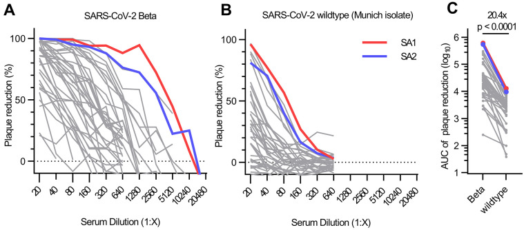
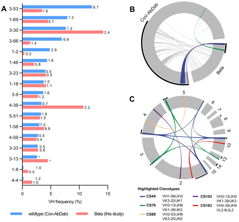
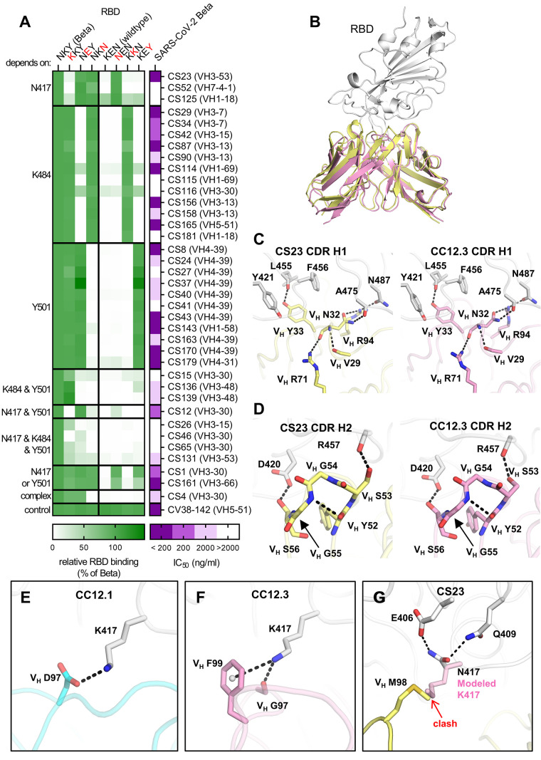
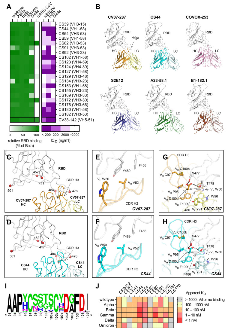
Severe acute respiratory syndrome coronavirus 2 (SARS-CoV-2) Beta variant of concern (VOC) resists neutralization by major classes of antibodies from COVID-19 patients and vaccinated individuals. In this study, serum of Beta-infected patients revealed reduced cross-neutralization of wild-type virus. From these patients, we isolated Beta-specific and cross-reactive receptor-binding domain (RBD) antibodies. The Beta-specificity results from recruitment of VOC-specific clonotypes and accommodation of mutations present in Beta and Omicron into a major antibody class that is normally sensitive to these mutations. The Beta-elicited cross-reactive antibodies share genetic and structural features with wild type-elicited antibodies, including a public VH1-58 clonotype that targets the RBD ridge. These findings advance our understanding of the antibody response to SARS-CoV-2 shaped by antigenic drift, with implications for design of next-generation vaccines and therapeutics.


Figures

Fig. 1.
Fig. 1.. Authentic virus neutralization of sera from individuals after infection with SARS-CoV-2 Beta.
(A and B) Neutralizing activity of sera of patients infected with SARS-CoV-2 Beta variant was measured using a plaque-reduction neutralization assay with the indicated authentic virus. Results are given as reduction of plaque number at indicated serum dilutions. Patients SA1 and SA2 mounted the strongest antibody response, which are highlighted in red and blue, respectively. Means of duplicate measurements are shown. Values below zero indicate no plaque reduction. (C) Change in neutralization activity against SARS-CoV-2 Beta and wildtype SARS-CoV-2 based on area under the curve (AUC) calculations from authentic virus PRNT curves [shown in (A) and (B)]. Mean fold change is indicated above the p value. Statistical analysis was performed using a Wilcoxon matched-pairs signed-rank test with two-tailed p value.
Fig. 2.
Fig. 2.. Germline gene usage and clonotype analysis of Beta-elicited antibodies.
(A) VH gene usage of 289 RBD Beta IgG mAbs from this study (red) is compared to 1037 wildtype RBD mAbs from 96 previously published studies (blue, CoV-AbDab) (17). Frequencies of mAbs encoded by each VH gene are shown as bars. Enrichment of indicated VH genes is compared to healthy individuals (31) with fold-enrichment shown as number next to bars. VH gene frequencies that were not reported in healthy individuals (31) are shown with asterisk (*). Only VH genes with a frequency of at least 2% in CoV-AbDab are shown and VH genes are ordered by frequency in CoV-AbDab. (B) Circos plot shows the relationship between 289 IgG mAbs from this study (Beta) and 1037 previously published human mAbs reactive to RBD (wildtype) from 96 studies (17). Interconnecting lines display clonotypes shared between both datasets, as defined by the usage of the same V and J gene on both Ig heavy and light chain. Thin black lines at the outer circle border indicate expanded clonotypes within the respective data set. (C) Circos plot displaying the 289 IgG mAbs from this study grouped per patient. Interconnecting colored lines indicate clonotypes found in more than one patient. Small black angles at the outer circle border indicate clonally expanded clones within one patient. [(B) and (C)] Colored interconnecting lines depict clonotypes found in more than one patient of our cohort.
Fig. 3.
Fig. 3.. Binding, neutralization and structures of Beta-specific antibodies.
(A) Neutralization of indicated Beta-specific mAbs against authentic Beta virus is shown in purple. Binding to single point mutant RBD constructs with the indicated amino-acid residues at positions 417, 484 and 501 is shown in green, normalized to RBD Beta. (B to G) Structural comparison of VH3-53 mAbs between Beta-specific CS23 and wildtype-specific CC12.1 and CC12.3. (B) CC12.3 and CS23 adopt the same binding mode. The crystal structure of CC12.3 (pink) in complex with RBD (wildtype) was superimposed onto CS23 (yellow) in complex with RBD (Beta). Only the variable domains of the antibodies are shown for clarity. A small local conformational difference was observed between CS23-bound RBD Beta and CC12.3-bound wildtype RBD (191 Cα, RMSD = 0.8 Å). (C and D) Comparison of the (C) CDR H1 (‘NY’ motif) and (D) CDR H2 (‘SGGS’ motif) between CS23 and CC12.3. (E to G) Structures of CDR H3 of (E) CC12.1, (F) CC12.3, and (G) CS23. A modeled side chain of K417 is shown as transparent pink sticks, which would be unfavorable for binding to CS23, where VH M98 occupies this pocket. Structures of CC12.1 (PDB 6XC3, cyan), CC12.3 (PDB 6XC4, pink), and CS23 (this study, yellow) are used throughout this figure, and the RBD is shown in white. Hydrogen bonds, salt bridges or cation-π bonds are represented by black dashed lines.
Fig. 4.
Fig. 4.. Characterization of cross-reactive mAbs and crystal structures of CV07-287 and CS44.
(A) Neutralization of cross-reactive antibodies against authentic Beta, Delta and wildtype virus is shown in purple. Binding to the indicated RBD constructs is shown in green, normalized to RBD Beta. (B) VH1-58 antibodies target SARS-CoV-2 RBD via the same binding mode. Crystal structures of CV07-287 in complex with wildtype RBD and CS44 in complex with RBD Beta are shown. COVA1-16 Fab that was used in the crystallization to form the crystal lattice is not shown for clarity. Structures of VH1-58 antibodies from other studies are shown for comparison, including COVOX-253 (PDB 7BEN), S2E12 (PDB 7K45), A23-58.1 (PDB 7LRT), and B1-182.1 (PDB 7MM0). All structures are shown in the same orientation, with the constant domains of the Fab omitted for clarity. The location of the ridge region of the RBD is indicated in the first panel. (C and D) Mutated residues in VOCs B.1.1.7 (Alpha), B.1.351 (Beta), B.1.617.2 (Delta), and P.1 (Gamma) variants are represented by red spheres. All of these residues are distant from VH1-58 antibodies (C) CV07-287 and (D) CS44, except for T478. The disulfide bond in each CDR H3 is shown as sticks. (E to H) Detailed interactions between the RBD and [€ and (G)] CV07-287, and [(F) and (H)] CS44, respectively. RBDs are shown in white, with heavy and light chains of CV07-287 in orange and yellow, and those of CS44 in cyan and light cyan, respectively. Interactions of CDR H2 are shown in € and (F), and those of CDR H3 are in (G) and (H). Hydrogen bonds are represented by black dashed lines. (I) Sequence logo of CDR H3 of VH1-58/VK3-20 antibodies. CDR H3 sequences of VH1-58/VK3-20 antibodies from COVID-19 patients (17) were aligned and analyzed with WebLogo. (J) Affinity of indicated Beta-elicited mAbs to RBD of indicated VOCs was determined by biolayer interferometry.

References

    1. Dejnirattisai W., Zhou D., Supasa P., Liu C., Mentzer A. J., Ginn H. M., Zhao Y., Duyvesteyn H. M. E., Tuekprakhon A., Nutalai R., Wang B., López-Camacho C., Slon-Campos J., Walter T. S., Skelly D., Costa Clemens S. A., Naveca F. G., Nascimento V., Nascimento F., Fernandes da Costa C., Resende P. C., Pauvolid-Correa A., Siqueira M. M., Dold C., Levin R., Dong T., Pollard A. J., Knight J. C., Crook D., Lambe T., Clutterbuck E., Bibi S., Flaxman A., Bittaye M., Belij-Rammerstorfer S., Gilbert S. C., Carroll M. W., Klenerman P., Barnes E., Dunachie S. J., Paterson N. G., Williams M. A., Hall D. R., Hulswit R. J. G., Bowden T. A., Fry E. E., Mongkolsapaya J., Ren J., Stuart D. I., Screaton G. R., Antibody evasion by the P.1 strain of SARS-CoV-2. Cell 184, 2939–2954.e9 (2021). 10.1016/j.cell.2021.03.055 - DOI - PMC - PubMed
    1. Wang P., Nair M. S., Liu L., Iketani S., Luo Y., Guo Y., Wang M., Yu J., Zhang B., Kwong P. D., Graham B. S., Mascola J. R., Chang J. Y., Yin M. T., Sobieszczyk M., Kyratsous C. A., Shapiro L., Sheng Z., Huang Y., Ho D. D., Antibody resistance of SARS-CoV-2 variants B.1.351 and B.1.1.7. Nature 593, 130–135 (2021). 10.1038/s41586-021-03398-2 - DOI - PubMed
    1. Hoffmann M., Arora P., Groß R., Seidel A., Hörnich B. F., Hahn A. S., Krüger N., Graichen L., Hofmann-Winkler H., Kempf A., Winkler M. S., Schulz S., Jäck H.-M., Jahrsdörfer B., Schrezenmeier H., Müller M., Kleger A., Münch J., Pöhlmann S., SARS-CoV-2 variants B.1.351 and P.1 escape from neutralizing antibodies. Cell 184, 2384–2393.e12 (2021). 10.1016/j.cell.2021.03.036 - DOI - PMC - PubMed
    1. Hoffmann M., Hofmann-Winkler H., Krüger N., Kempf A., Nehlmeier I., Graichen L., Arora P., Sidarovich A., Moldenhauer A.-S., Winkler M. S., Schulz S., Jäck H.-M., Stankov M. V., Behrens G. M. N., Pöhlmann S., SARS-CoV-2 variant B.1.617 is resistant to bamlanivimab and evades antibodies induced by infection and vaccination. Cell Rep. 36, 109415 (2021). 10.1016/j.celrep.2021.109415 - DOI - PMC - PubMed
    1. Planas D., Bruel T., Grzelak L., Guivel-Benhassine F., Staropoli I., Porrot F., Planchais C., Buchrieser J., Rajah M. M., Bishop E., Albert M., Donati F., Prot M., Behillil S., Enouf V., Maquart M., Smati-Lafarge M., Varon E., Schortgen F., Yahyaoui L., Gonzalez M., De Sèze J., Péré H., Veyer D., Sève A., Simon-Lorière E., Fafi-Kremer S., Stefic K., Mouquet H., Hocqueloux L., van der Werf S., Prazuck T., Schwartz O., Sensitivity of infectious SARS-CoV-2 B.1.1.7 and B.1.351 variants to neutralizing antibodies. Nat. Med. 27, 917–924 (2021). 10.1038/s41591-021-01318-5 - DOI - PubMed

Publication types

MeSH terms

Substances

Supplementary concepts