

Investigation of HoxB3 and Growth Factors Expression in Placentas of Various Gestational Ages
- PMID: 35076557
- PMCID: PMC8788416
- DOI: 10.3390/jdb10010002
Investigation of HoxB3 and Growth Factors Expression in Placentas of Various Gestational Ages
Abstract
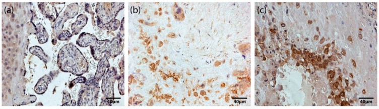
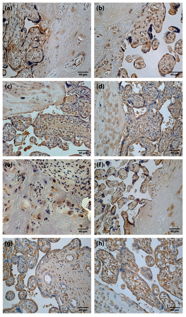
An evaluation of transforming growth factor beta (TGFβ), hepatocyte growth factor (HGF), basic fibroblast growth factor (FGF-2), fibroblast growth factors receptor 1 (FGFR1) and Hox-positive cells in the human placenta, and their correlation with gestational time at delivery and pregnancy outcomes, may provide not only a better understanding of the role of Hox genes and growth factors in human development, but also may be of clinical importance in reproductive medicine. This study analyzed the immunohistochemical identification of TGFβ, HGF, FGF-2, FGFR1 and HoxB3 in placentas of various gestational ages. We found few (+) TGFβ, moderate (++) FGF-2 and numerous (+++) HGF and FGFR1 positive structures. Occasional (0/+) to numerous (+++) HoxB3-positive structures were detected in different types of placental cells specifically, cytotrophoblasts, syncytiotrophoblast, extravillous trophoblasts, and Höfbauer cells. Correlating the appearance of HoxB3 staining in placentas with neonatal parameters, we found a statistically significant negative correlation with ponderal index (r = -0.323, p = 0.018) and positive correlation with neonate body length (r = 0.541, p = 0.046). The number of HoxB3-positive cells did not correlate with growth factors and gestational age, but with neonatal anthropometrical parameters, indicating the role of HoxB3 not only in placental development, but also in the longitudinal growth of the fetus. TGFβ and FGF-2 did not play a significant role in the development of the placenta beyond 22nd week of pregnancy, while HGF and FGFR1 immunoreactive cells increased with advancing gestation, indicating increasingly evolving maturation (growth, proliferation) of the placenta, especially in the third trimester.
Keywords: development; fetus; gene; growth; immunocytochemistry; intrauterine growth; microscopy; placenta.
Conflict of interest statement
The authors declare no conflict of interest.
Figures
Similar articles
-
Assessment of apoptosis and appearance of hepatocyte growth factor in placenta at different gestational ages: A cross-sectional study.Int J Reprod Biomed. 2021 Jul 27;19(6):505-514. doi: 10.18502/ijrm.v19i6.9372. eCollection 2021 Jun. Int J Reprod Biomed. 2021. PMID: 34401645 Free PMC article.
-
Fibroblast growth factor-2 and fibroblast growth factor receptor-1 mRNA expression and peptide localization in placentae from normal and diabetic pregnancies.Placenta. 1998 Mar-Apr;19(2-3):133-42. doi: 10.1016/s0143-4004(98)90001-7. Placenta. 1998. PMID: 9548179
-
Colocalization of acidic and basic fibroblast growth factor (FGF) in human placenta and the cellular effects of bFGF in trophoblast cell line JEG-3.Growth Factors. 1994;10(4):259-68. doi: 10.3109/08977199409010992. Growth Factors. 1994. PMID: 7528516
-
Placental expression of insulin-like growth factor-I, fibroblast growth factor-basic and neural cell adhesion molecule in pregnancies with small for gestational age fetuses.J Perinatol. 2008 Jul;28(7):468-74. doi: 10.1038/jp.2008.27. Epub 2008 Apr 24. J Perinatol. 2008. PMID: 18432248
-
Viral infection, proliferation, and hyperplasia of Hofbauer cells and absence of inflammation characterize the placental pathology of fetuses with congenital Zika virus infection.Arch Gynecol Obstet. 2017 Jun;295(6):1361-1368. doi: 10.1007/s00404-017-4361-5. Epub 2017 Apr 11. Arch Gynecol Obstet. 2017. PMID: 28396992 Free PMC article. Review.
Cited by
-
SIRT5 suppresses the trophoblast cell proliferation, invasion, and migration to promote preeclampsia via desuccinylating HOXB3.J Assist Reprod Genet. 2024 Oct;41(10):2759-2770. doi: 10.1007/s10815-024-03223-5. Epub 2024 Aug 15. J Assist Reprod Genet. 2024. PMID: 39145876
References
LinkOut - more resources
Full Text Sources
Miscellaneous
