Review
. 2022 Jan 13;8(1):200-228.
doi: 10.3390/tomography8010017.

MDCT Imaging of Non-Traumatic Thoracic Aortic Emergencies and Its Impact on Diagnosis and Management-A Reappraisal

Affiliations
Review

MDCT Imaging of Non-Traumatic Thoracic Aortic Emergencies and Its Impact on Diagnosis and Management-A Reappraisal

Tullio Valente et al. Tomography. .

Abstract

Non-traumatic thoracic aorta emergencies are associated with significant morbidity and mortality. Diseases of the intimomedial layers (aortic dissection and variants) have been grouped under the common term of acute aortic syndrome because they are life-threatening conditions clinically indistinguishable on presentation. Patients with aortic dissection may present with a wide variety of symptoms secondary to the pattern of dissection and end organ malperfusion. Other conditions may be seen in patients with acute symptoms, including ruptured and unstable thoracic aortic aneurysm, iatrogenic or infective pseudoaneurysms, aortic fistula, acute aortic thrombus/occlusive disease, and vasculitis. Imaging plays a pivotal role in the patient's management and care. In the emergency room, chest X-ray is the initial imaging test offering a screening evaluation for alternative common differential diagnoses and a preliminary assessment of the mediastinal dimensions. State-of-the-art multidetector computed tomography angiography (CTA) provides a widely available, rapid, replicable, noninvasive diagnostic imaging with sensitivity approaching 100%. It is an impressive tool in decision-making process with a deep impact on treatment including endovascular or open surgical or conservative treatment. Radiologists must be familiar with the spectrum of these entities to help triage patients appropriately and efficiently. Understanding the imaging findings and proper measurement techniques allow the radiologist to suggest the most appropriate next management step.

Keywords: TEVAR; acute aortic syndrome; aorta; aortic aneurysm; aortic dissection; aortic emergencies; computed tomography angiography; emergency CT; imaging of the aorta; management.


Conflict of interest statement

The authors declare no conflict of interest.

Figures

Figure 1
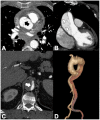
Figure 1
Computed tomography angiography (CTA) provides a rapid noninvasive diagnosis of the entire aorta including aberrant anatomy and pathology. (A) A 74-year-old man presenting in ED with 10 h of abrupt onset of acute chest pain, improving anemia, hypotension (blood pressure of 70/40 mm Hg in both upper extremities), and shortness of breath. CTA shows a right aortic arch and a pleural and extrapleural left hematoma from an aneurysm (asterisk) of a retroesophageal aberrant left subclavian artery (ALSA). (B) More caudal axial scan shows the ALSA aneurysm rupture site (arrow). Note the anomalous course of the left brachiocefalic vein posterior to the ascending aorta. (C) CTA-delayed phase shows contrast medium extravasation in the extrapleural hematoma (asterisk); note extrapleural fat sign (arrow). (D) 3D volume rendering oblique sagittal reformat image clearly shows the aneurysm (asterisk) coming immediately off the ALSA origin (arrow) from a Kommerell’s diverticulum (arrowhead).
Figure 2
Figure 2
Usefulness of CTA low-dose unenhanced phase in acute aortic syndrome. (A) Noncontrast CT axial scan shows dissection flap (arrow) and displaced intimal calcifications (arrowhead) in supra-aortic vessels extension of type A aortic dissection in a 79-year-old-man with acute neck and back pain. (B) Axial image from noncontrast CT shows a crescent-shaped high-density rind in the ascending aorta, a typical appearance of type A acute intramural hematoma (arrowheads). (C) Noncontrast CT axial scan shows a large bilateral pleural effusion with increased density in the dependent aspect of the collection (hematocrit sign), highly suggestive of hemothorax (asterisk) from rupture of the descending thoracic aorta aneurysm. (D) Axial image of aorta from noncontrast CT shows hemomediastinum tracking along the main and right pulmonary arteries and hyperattenuating dissection flap (arrowheads) in ascending aorta in a 68-year-old woman with ruptured type A dissection.
Figure 3
Figure 3
(A) Coronal MPR image of CTA in a 57-year-old man with chest pain and troponin negative shows coexistence of an acute intramural hematoma in the ascending aorta (type A IMH, arrowheads) and a dual-barrel dissection in the abdominal aorta (arrows). (B,C) Axial CTA images in another patient, showing the extension of the dissection flap from the proximal arch (arrow) to its distal segment (arrowheads) after only 19 s in the same CTA examination. Aortic dissection may be a very dynamic process.
Figure 4
Figure 4
Pretest clinical probability assessment according to Aortic Dissection Detection Risk Score (ADD-RS) and D-Dimer (DD). ADD-RS score is based on 12 risk factors organized in three categories (0, low risk; 1, intermediate risk; 2–3. high risk). BP = blood pressure. * Caution in patients with early presentation (≤2 h) or long-lasting symptoms (≥1 week) [28].
Figure 5
Figure 5
CTA findings in aortic dissection. (A) Axial CTA image showing intimomedial rupture sign in the ascending aorta (arrow) in type A dissection, indicating direction of the intimomedial entrance tear from true to false lumen. (B) Coronal CTA MPR image showing the intimomedial flap travelling parallel to the aorta long axis. Note the cobweb sign by thin, string-like filaments of the media layer in the false lumen (arrowheads). (C) Axial CTA image shows pressure competition between lumina and opacified true lumen collapse (arrow). (D) The different density of the TL (more intensely opacified due to faster flow) and FL in the early angiographic phase allows them to be clearly distinguished in the coronal VR reconstruction.
Figure 6
Figure 6
CTA findings in classic dual-barrel aortic dissection. (A) Axial CTA images showing the “beak sign” (arrowheads) in the false lumen because its wall and intimomedial flap usually form acute angles resembling a bird’s beak. In the radiological report, maximum aortic diameter and minimum and maximum aortic true lumen diameter should be included. (B) Sagittal oblique MPR image of non-A-non-B dissection with descending-entry and retrograde arch extension (arrow) in a 75-year-old male presenting with acute chest pain. (C) Axial CTA images. Static aortic dissection is seen as protrusion of the intimal flap (arrow) into the ostium of the affected branch vessel causing subsequent partial or total thrombosis of the branch vessel with resulting perfusion impairments. Note left kidney partial infarct (arrowheads). (D) CTA oblique coronal VR reconstruction image shows a three lumina aortic dissection.
Figure 7
Figure 7
(A) Axial CTA image demonstrates thickening of the main pulmonary arterial wall (arrowheads) indicating IMH of the PA in ruptured Type A AD. (B) Axial CTA image shows entry tear (arrow) in the arch convexity in a 53-year-old man with severe chest pain. (C) 3D-VR parasagittal reconstruction image shows localized 4-vessels arch dissection (arrow) without supra-aortic trunks involvement. (D) 3D-VR parasagittal reconstruction image shows a non-A-non-B dissection with arch-entry and anterograde descending aorta involvement.
Figure 8
Figure 8
CT features of acute type B IMH in a 62-year-old man with abrupt chest pain and Type B IMH at baseline CTA examination. (A) Axial low-dose unenhanced CT image shows hyperdense crescent sign of the aortic wall (arrow). (B) Axial arterial phase CTA image in the same patient shows decreased diameter of the aortic lumen and the smooth luminal-wall interface. (C) Sagittal reconstruction CTA image shows decreased diameter of the aortic lumen (arrow) and a relative constant circumferential relationship with the wall.
Figure 9
Figure 9
Unstable Type A IMH complicated by ulcer-like projection (ULP). (A) Coronal oblique CTA shows type A IMH (arrowheads) in a 68-year-old man with hypertension and chest pain. The total aortic diameter was less than 50 mm and the IMH thickness less than 11 mm; he underwent medical management initially. (B) Three-day follow-up coronal oblique CTA image shows disease progression by an ULP due to opening of intimal tear (arrow). (C) 3D-volume-rendered (VR) reconstruction confirms ULP (arrow).
Figure 10
Figure 10
CTA features of Svensson’s Class 4 (PAU) and Class 3 (limited intimal tear) variants. (A) Axial unenhanced CT image shows aortic arch high-density intramural hematoma (IMH) in a 74-year-old male with acute neck and chest pain (arrowheads). (B) CTA axial scan in the same patient shows a flat penetrating ulcer (arrow) with associated IMH in the arch convexity. (C) CTA axial image shows a linear filling defect with subtle undermined edges (arrow) and eccentric medial one-sided bulge of the ascending aorta (arrowhead) without a clear intimomeadial flap or false lumen in a 58-year-old male with sudden onset of chest and back pain. (D) 3D-VR oblique coronal reconstruction confirms the eccentric one-sided saccular aortic bulging (‘‘mushroom cap’’ morphology) of class 3 dissection (arrow).
Figure 11
Figure 11
CTA of thoracic aorta aneurysms. (A) Marfan syndrome, a multisystem connective tissue disease caused by a defect in the protein fibrillin 1, and annuloaortic ectasia in a 42-year-old man. Sagittal MIP reconstruction image shows a proximal dilatation of the aortic anulus (1), sinuses of Valsalva (2) along with effacement of the STJ (4) producing a pear-shaped ascending aorta (3). (B) Axial CTA image shows sinuses ectasia in the same patient. In Marfan syndrome, a cut-off value of 5 cm of the ascending aorta diameter is recommended for surgical repair. (C) Axial CTA image shows a 12 cm ascending aorta aneurysm (asterisk) and a large mediastinal effusion. The risk of rupture of TAAs increases with size of the aneurysm according to Laplace’s law. (D) Axial CTA image shows a ruptured atherosclerotic aneurysm of the descending thoracic aorta. Note the high-attenuation fluid in the right pleural space, representing acute hemothorax (asterisk), and contrast medium extravasation from the aortic lumen (arrow).
Figure 12
Figure 12
CTA findings in impending rupture of the thoracic aorta aneurysms. (A) Missing calcium sign (arrowhead). (B) Thrombus fissuration (arrow). (C) Hyperattenuating crescent sign, periaortic stranding sign, and contour irregularity of the aneurysm. (D) Periaortic fat stranding blurs a focal area of the mediastinum and extends to the chest wall representing an inflammatory process (arrowheads) [111].
Figure 13
Figure 13
CT evaluation and CT-PET in a 68-year-old male patient with an aortoesophageal fistula (AEF) after 28 days from TEVAR. (A,B) CTA evaluation for the onset of fever showing disappearance of thoracic descending aortic wall calcification (arrow), distended esophagus (curved arrow), and appearance of an ectopic small air bubble in the native aneurysmal aortic lumen (arrowhead). (C) Coronal oblique MPR image better shows a significant amount of air in the native aortic lumen (arrowheads) due to probable AEF. (D) Fused FDG PET-CT axial image shows multiple foci of increased FDG uptake both in the aneurysm wall (arrowheads) both in the mediastinum (arrow) suggestive of infected thoracic aortic aneurysm and TEVAR endograft.
Figure 14
Figure 14
CTA of aortitis and periaortitis by Salmonella species. A 68-year-old man with chills, high temperature, and upper back pain radiating to anterior chest. (A) Unenhanced CTA axial image shows marginal periaortic tissue (asterisk) at aortic isthmus level. (B) Axial CTA late phase image also shows a focal outpouching (curved arrow) of contrast material emanating from the isthmus. (C,D) Axial arterial and delayed-phase CTA images obtained 7 days later show that contrast outpouching was considerably larger (curved arrows), a finding referable to rapidly growing focal mycotic pseudoaneurysm, and the patient underwent endovascular aortic repair and intravenous antibiotic therapy.
Figure 15
Figure 15
CTA of acute diffuse aortic atheromatous embolization in a 46-year-old COVID-19 pneumonia patient with acute cerebral vascular event. (A) CTA axial scan shows extensive and irregular atheroma of the ascending aorta (arrow). (B) Coronal MIP reconstruction image confirms ascending aorta filling defect (arrow). (C) In the same patient, CTA axial scan shows extensive irregular atheroma of the transverse aortic arch. (D) Sagittal MIP reconstruction image confirms extensive arch atheroma and friable floating thrombi in descending thoracic aorta (arrowheads).

References

    1. Jen J.P., Malik A., Lewis G., Holloway B. Non-traumatic thoracic aortic emergencies: Imaging diagnosis and management. Br. J. Hosp. Med. 2020;81:1–12. doi: 10.12968/hmed.2020.0344. - DOI - PubMed
    1. Voitle E., Hofmann W., Cejna M. Aortic emergencies-diagnosis and treatment: A pictorial review. Insights Imaging. 2015;6:17–32. doi: 10.1007/s13244-014-0380-y. - DOI - PMC - PubMed
    1. Lai V., Tsang W.K., Chan W.C., Yeung T.W. Diagnostic accuracy of mediastinal width measurement on posteroanterior and anteroposterior chest radiographs in the depiction of acute nontraumatic thoracic aortic dissection. Emerg. Radiol. 2012;19:309–315. doi: 10.1007/s10140-012-1034-3. - DOI - PMC - PubMed
    1. von Kodolitsch Y., Nienaber C.A., Dieckmann C., Schwartz A.G., Hofmann T., Brekenfeld C. Chest radiography for the diagnosis of acute aortic syndrome. Am. J. Med. 2004;116:73–77. doi: 10.1016/j.amjmed.2003.08.030. - DOI - PubMed
    1. Chawla A., Rajendran S., Yung W.H., Babu S.B., Peh W.C. Chest radiography in acute aortic syndrome: Pearls and pitfalls. Emerg. Radiol. 2016;23:405–412. doi: 10.1007/s10140-016-1415-0. - DOI - PubMed

LinkOut - more resources