Review
. 2022 Feb 28;50(1):55-69.
doi: 10.1042/BST20210819.

Molecular regulation of autophagosome formation

Affiliations
Review

Molecular regulation of autophagosome formation

Yan Hu et al. Biochem Soc Trans. .

Abstract

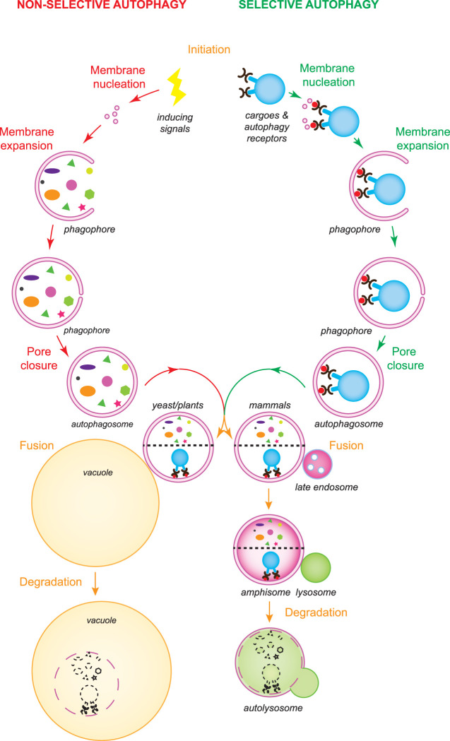
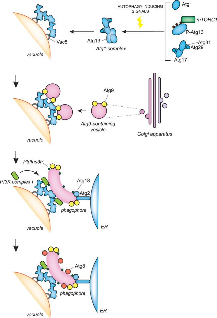
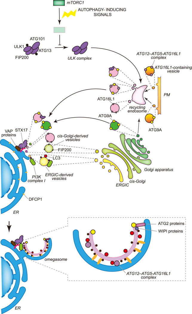
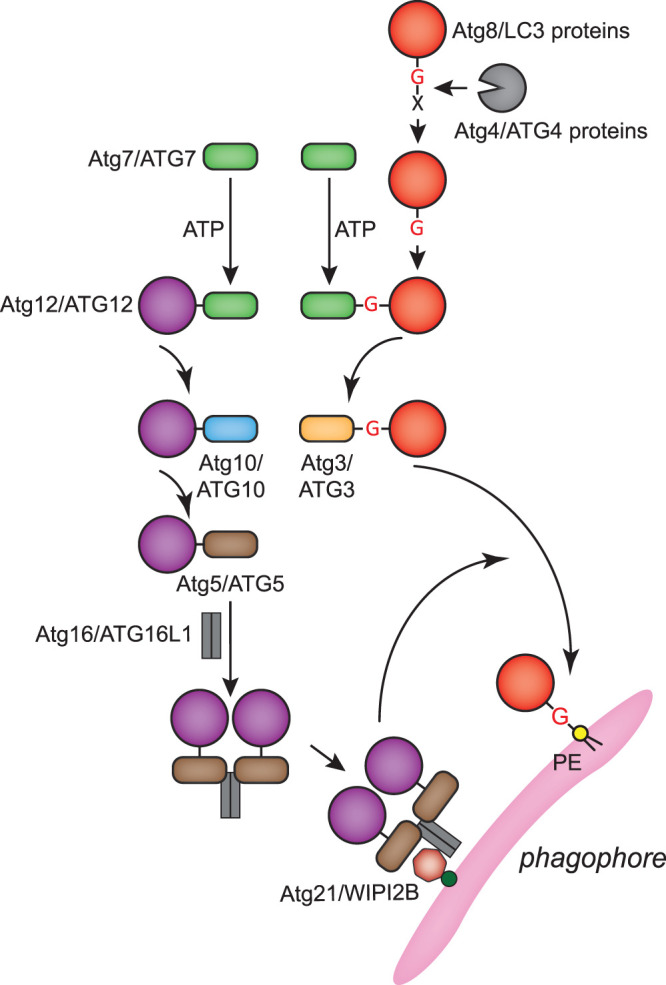
Macroautophagy, hereafter autophagy, is a degradative process conserved among eukaryotes, which is essential to maintain cellular homeostasis. Defects in autophagy lead to numerous human diseases, including various types of cancer and neurodegenerative disorders. The hallmark of autophagy is the de novo formation of autophagosomes, which are double-membrane vesicles that sequester and deliver cytoplasmic materials to lysosomes/vacuoles for degradation. The mechanism of autophagosome biogenesis entered a molecular era with the identification of autophagy-related (ATG) proteins. Although there are many unanswered questions and aspects that have raised some controversies, enormous advances have been done in our understanding of the process of autophagy in recent years. In this review, we describe the current knowledge about the molecular regulation of autophagosome formation, with a particular focus on budding yeast and mammalian cells.

Keywords: ATG proteins; autophagy; degradation; lysosomes; phagophore; sequestration.


Conflict of interest statement

The authors declare that there are no competing interests associated with the manuscript.

Figures

Figure 1.
Figure 1.. Overview of the autophagy process.
There are two types of autophagy: non-selective and selective autophagy. While non-selective bulk autophagy is induced by stress and other signals, selective autophagy is triggered by cargoes that must be turned over and autophagy receptors (black pincers) are central in this induction event. The fusion of vesicles, probably of different origins, leads to the phagophore nucleation. Subsequently, the expansion of the phagophore by the acquisition of extra lipids and the closure of the final pore lead to the formation of an autophagosome. The intracellular material that has to be recycled is sequestered during autophagosome biogenesis. By interacting with the Atg8/LC3-PE pool (red circles) in the inner surface of the expanding phagophore, the autophagosome receptors guide the exclusive sequestration of the cargoes targeted to degradation. Complete autophagosomes fuse with vacuoles in yeast and plants, and with lysosomes in mammals, delivering their cargo into the acidic lumen of these organelles. Autophagosomes can also fuse with late endosomes in plants and mammals, before fusing with vacuoles and lysosomes, respectively. Vacuolar and lysosomal resident hydrolases degrade the delivered cargoes into basic metabolites, which are then transported into the cytoplasm for recycling.
Figure 2.
Figure 2.. The molecular mechanisms of the early stages of autophagosome biogenesis during bulk autophagy in S. cerevisiae
In S. cerevisiae, autophagosome biogenesis takes place at the PAS, which is adjacent to both the vacuole and ER. Autophagy induction by nutrient starvation leads to an inhibition of TORC1 activity and the concomitant dephosphorylation of Atg13 promotes the assembly of the Atg1 kinase complex, which comprises Atg1 and the Atg17–Atg31–Atg29 subcomplex. Atg1 kinase complexes assemble into a supra-structure, probably by liquid phase separation, which is anchored onto the vacuole via binding to Vac8. Atg9-containing vesicles, which are derived from the Golgi apparatus, are recruited onto those supra-structures and by possibly fusing together and with vesicles of other origins, generate the phagophore. The PI3K complex I is recruited to the PAS via an interaction with Vac8 and produces PtdIns3P on the phagophore membrane. This phosphoinositide is involved in the recruitment and assembly of the Atg2–Atg18 complex, which probably transfers lipids from the ER to the phagophore for its expansion. Atg2 binds Atg9 and PtdIns3P on the phagophore and one or more unknown proteins on the ER, while Atg18 associates to the phagophore by interacting with Atg2 and PtdIns3P. Another PtdIns3P-binding protein, Atg21, interacts with Atg16 to recruit the Atg12–Atg5–Atg16 complex, which is crucial for the conjugation of Atg8 to the PE present in the phagophore (see also Figure 4).
Figure 3.
Figure 3.. The molecular mechanisms of the early stages of autophagosome biogenesis during bulk autophagy in mammals.
In mammalian cells multiple autophagosomes are simultaneously formed adjacently to the ER, in specific regions known as omegasomes, in which DFCP1 concentrates. The activity of the ULK kinase complex, which is constitutively formed by ULK1 or ULK2, ATG13, FIP200 and ATG101, is inhibited for example by mTORC1 in nutrient-rich conditions through the phosphorylation of ULK1 and ATG13. Nutrient deprivation leads to the inhibition of mTORC1 and the concomitant dephosphorylation of ULK1 and ATG13. Active ULK1 kinase complex associates to the recycling endosomes via an unknown mechanism and exits in vesicles that also contain ATG9A and/or the ATG12–ATG5–ATG16L1 complex. ATG9A reaches the recycling endosomes from either the Golgi apparatus or the plasma membrane (PM) by vesicular traffic, while ATG16L1 only from the PM. Recycling endosome-derived vesicles relocalize in the proximity of the ER, possibly through the binding of ULK1 and FIP200 with the ER transmembrane proteins VAPA and VAPB or STX17. The generation of the phagophore very likely involves the fusion of vesicles from different origins, including ATG9A-containing vesicles generated at the Golgi apparatus, COPII-coated vesicles derived from the ERGIC and the HyPAS. The HyPAS is a compartment formed through the fusion between ATG16L1-containing endosomal membranes and cis-Golgi-derived FIP200-positive vesicles, which is mediated by STX17 and its interactors SERCA2, E-SYT2 and SIGMAR1. The lipidation of LC3 already begins on these COPII-coated vesicles and relies on PtdIns3P, which is produced by the PI3K complex I. As a result, transport via COPII-coated vesicles is a possible mechanism for the localization of the PI3K complex I to the nascent phagophore. PtdIns3P is involved in the assembly of the complexes formed by ATG2 and WIPI proteins. The complexes formed by the ATG2 and WIPI proteins probably transfer lipids from the ER to the phagophore. Synthesis of PtdIns3P on the phagophore also leads to the association of ER-localized and PtdIns3P-binding DFCP1, which permits the generation of the characteristic omegasomes. Contrary to yeast, the distribution of the ATG proteins on the phagophore in mammalian cells is unknown and therefore what drawn in the figure is a speculative representation.
Figure 4.
Figure 4.. Atg8/LC3 and Atg12/ATG12 conjugation systems.
Atg12/ATG12 and Atg8/LC3 proteins are ubiquitin-like proteins. The conjugation systems of these two proteins are interconnected and they ultimately covalently link Atg8/LC3 proteins to the PE present in the phagophore membrane during autophagy. Amino acids at the C-terminus of Atg8/LC3 proteins (indicated with a X in the draw) are post-translationally cleaved by the Atg4/ATG4 proteases to expose a C-terminal glycine (G). Processed Atg8/LC3 proteins are activated and finally conjugated to the amino group of PE through the sequential action of the E1 enzyme Atg7/ATG7 and the E2 enzyme Atg3/ATG3. The last step of this reaction is mediated by the Atg12–Atg5–Atg16/ATG12–ATG5–ATG16L1 complex, which is formed by the Atg12/ATG12 conjugation system and acts as an E3 enzyme. The Atg12/ATG12 conjugation system involves the sequential action of Atg7/ATG7 and the E2 enzyme Atg10/ATG10, and covalently links Atg12/ATG12 to Atg5/ATG5. The Atg12/ATG12–Atg5/ATG5 conjugate binds to Atg16/ATG16L1, which subsequently multimerizes to form the Atg12–Atg5–Atg16/ATG12–ATG5–ATG16L1 complex. This complex associates with the phagophore via interaction with both Atg21/WIPI2B and PtdIns3P.

Similar articles

Cited by

References

    1. Lahiri, V., Hawkins, W.D. and Klionsky, D.J. (2019) Watch what you (self-) eat: autophagic mechanisms that modulate metabolism. Cell Metab. 29, 803–826 10.1016/j.cmet.2019.03.003 - DOI - PMC - PubMed
    1. Mizushima, N. and Levine, B. (2020) Autophagy in human diseases. N. Engl. J. Med. 383, 1564–1576 10.1056/NEJMra2022774 - DOI - PubMed
    1. Yang, Z. and Klionsky, D.J. (2010) Eaten alive: a history of macroautophagy. Nat. Cell Biol. 12, 814–822 10.1038/ncb0910-814 - DOI - PMC - PubMed
    1. Klionsky, D.J., Cregg, J.M., Dunn, Jr., W.A., Emr, S.D., Sakai, Y., Sandoval, I.V.et al. (2003) A unified nomenclature for yeast autophagy-related genes. Dev. Cell 5, 539–545 10.1016/s1534-5807(03)00296-x - DOI - PubMed
    1. Kirkin, V. and Rogov, V.V. (2019) A diversity of selective autophagy receptors determines the specificity of the autophagy pathway. Mol. Cell 76, 268–285 10.1016/j.molcel.2019.09.005 - DOI - PubMed

Publication types

Substances