. 2022 May;53(5):1606-1614.
doi: 10.1161/STROKEAHA.121.036749. Epub 2022 Jan 26.

Multimodal Neural and Behavioral Data Predict Response to Rehabilitation in Chronic Poststroke Aphasia

Affiliations

Multimodal Neural and Behavioral Data Predict Response to Rehabilitation in Chronic Poststroke Aphasia

Anne Billot et al. Stroke. 2022 May.

Abstract

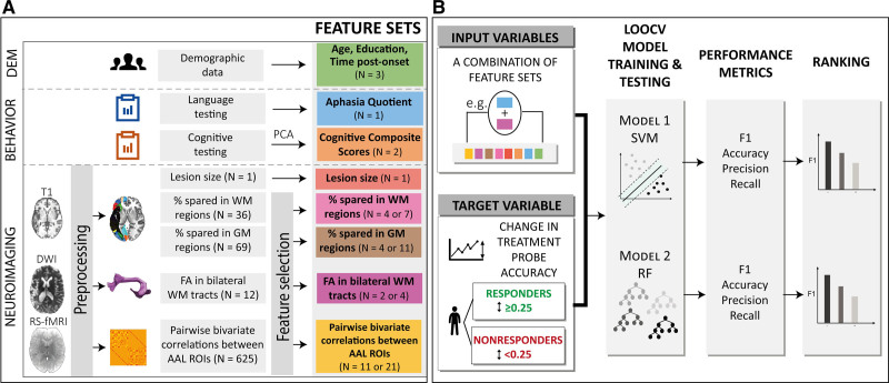
Background: Poststroke recovery depends on multiple factors and varies greatly across individuals. Using machine learning models, this study investigated the independent and complementary prognostic role of different patient-related factors in predicting response to language rehabilitation after a stroke.

Methods: Fifty-five individuals with chronic poststroke aphasia underwent a battery of standardized assessments and structural and functional magnetic resonance imaging scans, and received 12 weeks of language treatment. Support vector machine and random forest models were constructed to predict responsiveness to treatment using pretreatment behavioral, demographic, and structural and functional neuroimaging data.

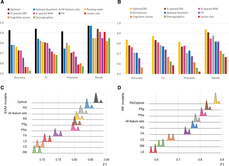
Results: The best prediction performance was achieved by a support vector machine model trained on aphasia severity, demographics, measures of anatomic integrity and resting-state functional connectivity (F1=0.94). This model resulted in a significantly superior prediction performance compared with support vector machine models trained on all feature sets (F1=0.82, P<0.001) or a single feature set (F1 range=0.68-0.84, P<0.001). Across random forest models, training on resting-state functional magnetic resonance imaging connectivity data yielded the best F1 score (F1=0.87).

Conclusions: While behavioral, multimodal neuroimaging data and demographic information carry complementary information in predicting response to rehabilitation in chronic poststroke aphasia, functional connectivity of the brain at rest after stroke is a particularly important predictor of responsiveness to treatment, both alone and combined with other patient-related factors.

Keywords: aphasia; language; machine learning; magnetic resonance imaging; neuroimaging; rehabilitation.


Figures

Figure 1.
Figure 1.
Methodological framework of the study. A, Behavioral, demographic, and neuroimaging data were collected before the commencement of the treatment. Neuroimaging data were preprocessed and feature selection was performed on feature sets with a high number of variables. B, All combinations of feature sets (N=255) were tested as input to the support vector machine (SVM) and random forest (RF) models to classify participants into responders and nonresponders. F1 score was used to rank models’ performance. AAL indicates Automated Anatomical Labeling atlas; DEM, demographics; DWI, diffusion weighted-imaging; FA, fractional anisotropy; GM, gray matter; LOOCV, leave-one-out cross-validation; PCA, principal component analysis; ROI, region of interest; RS-fMRI, resting-state functional MRI; and WM, white matter.
Figure 2.
Figure 2.
Lesion overlay for all participants. Z coordinates: −40 −30 −20 −10 0 10 20 30 40.
Figure 3.
Figure 3.
Change in accuracy on the treatment probes with accuracy measured as a percentage. Participants were classified into responders (R) and nonresponders (NR) to treatment based on a cutoff at 0.25. The different shades correspond to each participant’s site: Boston University (BU; dark), Johns Hopkins University (JHU; dark gray), and Northwestern University (NU; light gray).
Figure 4.
Figure 4.
Resting-state functional connectivity features. Red dot and lines represent functional regions and connections selected after feature selection and included in the machine learning models. Blue dots represent regions excluded from the analyses after feature selection. ACC indicates anterior cingulate cortex; AG, angular gyrus; FUS, fusiform gyrus; IFG, inferior frontal gyrus; INS, insula; IPG, inferior parietal gyrus; ITG, inferior temporal gyrus; L, left; midTP, middle temporal pole; MTG, middle temporal gyrus; orb, pars orbitalis; PCC, posterior cingulate gyrus; PCG, precentral gyrus; pre, pregenual; R, right; ROI, region of interest; SFG, superior frontal gyrus; STG, superior temporal gyrus; supTP, superior temporal pole; SMA, supplementary motor area; sub, subgenual; SMG, supramarginal; SOG, superior occipital gyrus; SPG, superior parietal gyrus; and tri, pars triangularis.
Figure 5.
Figure 5.
Predictive performance for support vector machine (SVM) and random forest (RF) models trained on a single feature set, all feature sets or the optimal combination of feature sets (aphasia quotient [AQ], demographics [DM], fractional anisotropy [FA], percentage spared in gray matter regions [PSg], resting-state [RS] for SVM and RS for RF). F1 scores of models trained on a single feature set were significantly lower than the models trained on the optimal combination of feature sets (P<0.001). A, Performance of SVM models on all 55 samples; (B) performance of RF models on all 55 samples; (C) distribution (kernel density estimation in R with automatic bandwidth selection) of SVM F1 scores computed from all 55 subsets of 54 samples each; and (D) distribution (kernel density estimation in R with automatic bandwidth selection) of RF F1 scores computed from all 55 subsets of 54 samples each.

References

    1. Adamson J, Beswick A, Ebrahim S. Is stroke the most common cause of disability? J Stroke Cerebrovasc Dis. 2004;13:171–177. doi: 10.1016/j.jstrokecerebrovasdis.2004.06.003 - PubMed
    1. Daniel K, Wolfe CD, Busch MA, McKevitt C. What are the social consequences of stroke for working-aged adults? A systematic review. Stroke. 2009;40:e431–e440. doi: 10.1161/STROKEAHA.108.534487 - PubMed
    1. Kauhanen ML, Korpelainen JT, Hiltunen P, Määttä R, Mononen H, Brusin E, Sotaniemi KA, Myllylä VV. Aphasia, depression, and non-verbal cognitive impairment in ischaemic stroke. Cerebrovasc Dis. 2000;10:455–461. doi: 10.1159/000016107 - PubMed
    1. Wilson SM, Eriksson DK, Brandt TH, Schneck SM, Lucanie JM, Burchfield AS, Charney S, Quillen IA, de Riesthal M, Kirshner HS, et al. . Patterns of recovery from aphasia in the first 2 weeks after stroke. J Speech Lang Hear Res. 2019;62:723–732. doi: 10.1044/2018_JSLHR-L-18-0254 - PMC - PubMed
    1. Watila MM, Balarabe SA. Factors predicting post-stroke aphasia recovery. J Neurol Sci. 2015;352:12–18. doi: 10.1016/j.jns.2015.03.020 - PubMed

Publication types