. 2022 Feb 8;119(6):e2113874119.
doi: 10.1073/pnas.2113874119.

Extracellular vimentin is an attachment factor that facilitates SARS-CoV-2 entry into human endothelial cells

Affiliations

Extracellular vimentin is an attachment factor that facilitates SARS-CoV-2 entry into human endothelial cells

Razie Amraei et al. Proc Natl Acad Sci U S A. .

Abstract

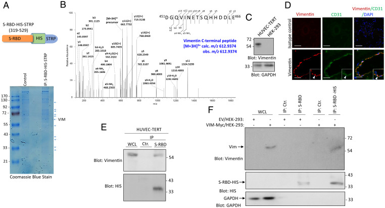
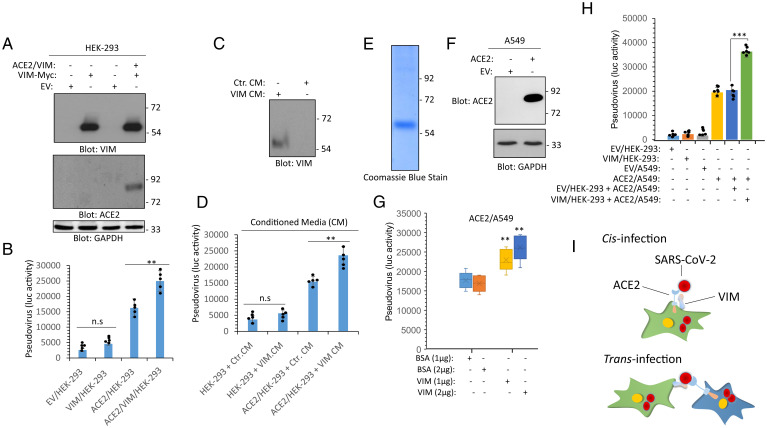
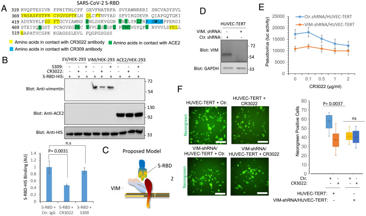
SARS-CoV-2 entry into host cells is a crucial step for virus tropism, transmission, and pathogenesis. Angiotensin-converting enzyme 2 (ACE2) has been identified as the primary entry receptor for SARS-CoV-2; however, the possible involvement of other cellular components in the viral entry has not yet been fully elucidated. Here we describe the identification of vimentin (VIM), an intermediate filament protein widely expressed in cells of mesenchymal origin, as an important attachment factor for SARS-CoV-2 on human endothelial cells. Using liquid chromatography-tandem mass spectrometry, we identified VIM as a protein that binds to the SARS-CoV-2 spike (S) protein. We showed that the S-protein receptor binding domain (RBD) is sufficient for S-protein interaction with VIM. Further analysis revealed that extracellular VIM binds to SARS-CoV-2 S-protein and facilitates SARS-CoV-2 infection, as determined by entry assays performed with pseudotyped viruses expressing S and with infectious SARS-CoV-2. Coexpression of VIM with ACE2 increased SARS-CoV-2 entry in HEK-293 cells, and shRNA-mediated knockdown of VIM significantly reduced SARS-CoV-2 infection of human endothelial cells. Moreover, incubation of A549 cells expressing ACE2 with purified VIM increased pseudotyped SARS-CoV-2-S entry. CR3022 antibody, which recognizes a distinct epitope on SARS-CoV-2-S-RBD without interfering with the binding of the spike with ACE2, inhibited the binding of VIM with CoV-2 S-RBD, and neutralized viral entry in human endothelial cells, suggesting a key role for VIM in SARS-CoV-2 infection of endothelial cells. This work provides insight into the pathogenesis of COVID-19 linked to the vascular system, with implications for the development of therapeutics and vaccines.

Keywords: ACE2; SARS-CoV-2; endothelial cells; vimentin; viral entry.


Conflict of interest statement

The authors declare no competing interest.

Figures

Fig. 1.
Fig. 1.
Identification of VIM as a SARS-CoV-2 S binding protein. (A) Schematic of SARS-CoV-2 S-RBD extended with a HIS and STRP. WCL from HUVEC-TERT cells were incubated with S-RBD-HIS-STRP-Ni-IMAG beads or Ni-IMAG beads alone and the captured proteins were resolved on SDS/PAGE and stained with Coomassie blue. The protein bands were cut out, subjected to in-gel digestion with trypsin, and the resulting peptide mixture was analyzed by LC-MS/MS. (B) HCD MS/MS spectrum is assigned to the C-terminal peptide of VIM, 451DGQVINETSQHHDDLE466 precursor [M + 3H]3+ calc. m/z 612.9374 obs. m/z 612.9374. Symbols: bn = N-terminal fragments; yn = C-terminal fragments. Peptide assignments were made by comparison to the Uniprot human database plus the sequence for the recombinant SARS-CoV-2 S-protein, using the Andromeda search engine with MaxQuant v1.6.14, and were checked manually. Seven VIM peptides, representing 14.4% of the protein amino acid sequence, were assigned with high confidence in the LC-MS/MS dataset. (C) Western blot analysis of VIM expression in HUVEC-TERT and control HEK-293 cells. The same membrane was blotted for GAPDH as a protein loading control. (D) PFA fixed human lung tissue was subjected to immunofluorescence staining. The tissue was stained with an anti-CD31 (endothelial marker) and anti-VIM antibodies. Image magnification: 50 µM. (E) WCL from HUVEC-TERT cells were incubated with S-RBD-HIS-STRP-Ni-IMAG beads or Ni-IMAG beads alone. The captured proteins were resolved on SDS/PAGE and blotted with an anti-VIM antibody. The same membrane was blotted with anti-HIS for S-RBD-HIS-STRP. (F) WCL from HEK-293 cells expressing control EV or VIM-Myc. The whole lysates were incubated with S-RBD-HIS-STRP-Ni-IMAG beads or Ni-IMAG beads alone and after extensive washing, and the captured proteins were resolved on SDS-PAGE and blotted with anti-VIM antibody. The same membrane was reblotted with anti-HIS for S-RBD-HIS-STRP or with GAPDH as the protein loading control.
Fig. 2.
Fig. 2.
VIM intermediate filament is present on the extracellular surface and acts as an attachment factor for SARS-CoV-2. (A) HEK-293 cells were transfected with plasmids expressing mCherry or VIM-mCherry. Forty-eight hours after transfection, cells were fixed and the slides were viewed under a fluorescence microscope. Cell nuclei were stained with DAPI (blue). (Scale bars, 50 µm.) (B) HEK-293 cells expressing VIM were stained with an anti-VIM antibody and DAPI. The slides were viewed under a confocal microscope. (Scale bar, 50 µm; Inset, 50 µm.) (C) CM from HEK-293 cells transfected with an EV or a plasmid expressing VIM-mCherry were concentrated, resolved by SDS/PAGE, and subjected to Western blot analysis using an anti-VIM antibody. (D) HEK-293 cells expressing EV or VIM-Myc were subjected to cell surface biotinylation. Cells were lysed and cell lysates were immunoprecipitated with an anti-VIM antibody followed by Western blot analysis using Streptavidin antibody. The same membrane was stripped off the antibody and reblotted with anti-Myc antibody. (E) HEK-293 cells expressing EV, VIM or ACE2 were plated in 96-well plates (2.5 × 10⁵ cells per well, five wells per group). After overnight incubation, cells were treated with mock or SARS-CoV-2 S pseudovirus-luc and luciferase activity was measured after additional 24 h. (F) HEK-293 cells expressing EV, VIM, or ACE2 were plated in 96-well plates as described for C. Cells were treated with mock or SARS-CoV-2 S pseudovirus-GFP. After 48 h, cells were subjected to live cell imaging and pictures were taken from each well from random fields. GFP+ cells were counted. Graph is quantification of GFP+ cells (five well per group). (G) HEK-293 cells expressing EV, VIM-Myc, or ACE2 were seeded in 96-well plates (triplicate per group) and infected with SARS-CoV-2-mNG at an MOI of 2. After 24 h, cells were fixed in 10% neutral buffered formalin followed by staining with DAPI. Cells were viewed under a Nikon deconvolution fluorescence microscope and pictures were taken from random fields. Graph shows quantification of SARS-CoV-2-mNG+ cells (triplicates well per group, from two independent experiments). (Scale bars, 50 µm.)
Fig. 3.
Fig. 3.
VIM binds to ACE2 and increases the binding of SARS-CoV-2 S-protein to ACE2. (A) WCL from HEK293 cells expressing an EV, VIM-Myc or coexpressing VIM-Myc with ACE2 were subjected to Western blot analysis using anti-VIM, anti-ACE2, or GAPDH antibodies. (B) HEK293 cells expressing EV, VIM-Myc, ACE2, or coexpressing VIM-Myc with ACE2 were plated in 96-well plates (2.5 × 10⁵ cells per well, five wells per group). After overnight incubation, cells were treated with mock or SARS-CoV-2 S pseudovirus-luc. After 24 h. cells were lysed and luciferase activity was measured. (C) Western blot analysis of CM from control HEK-293 cells or HEK-293 cells expressing VIM. (D) HEK-293 cells expressing EV or ACE2 were seeded in 96-well plates (2.5 × 10⁵ cells per well, five wells per group). Cells were treated with control CM (Ctr. CM) or CM from HEK-293 cells expressing VIM. After 1-h incubation, the media was removed and the cells were subjected to virus entry assay using SARS-CoV-2 S pseudovirus-luc. After 24 h, cells were lysed and luciferase activity was determined. cells expressing EV or VIM or ACE2 were plated in 96-well plates (2.5 × 10⁵ cells per well, five wells per group). After overnight incubation, cells were preincubated with purified VIM (1 or 2 µg per well) or the control vehicle (BSA). After 30 min, cells were washed with PBS to remove the unbound VIM. Cells then were treated with mock or SARS-CoV-2 S pseudovirus-luc and luciferase activity was measured after an additional 24 h. **P < 0.01, ***P < 0.001; n.s., not significant.
Fig. 4.
Fig. 4.
Extracellular VIM facilitates SARS-CoV-2 entry into human cells. (A) HEK-293 cells expressing ACE2 and VIM-Myc were costained with anti-ACE2, anti-VIM and DAPI. The slides were viewed by confocal microscopy. White arrowheads show colocalization of VIM with ACE2. (Scale bars, 50 µM.) (B) WCL from HEK-293 cells expressing control EV or coexpressing VIM-Myc with ACE2 were subjected to a coimmunoprecipitation assay using anti-ACE2 antibody. The immunoprecipitated proteins were analyzed by Western blot analysis using anti-Myc antibody for VIM. The same membrane was also probed for ACE2 and GAPDH. (C) Different concentrations of WCL from HEK-293 cells expressing EV, VIM, ACE2, or coexpressing VIM with ACE2 were blotted on the PVDF membrane. The membranes after blocking with BSA were incubated with S-RBD-HIS-STRP (1 µg/mL), followed with immunoblotting with anti-HIS antibody. Quantification of the blots is shown. (D) A 96-well plate coated with soluble ACE2 was incubated with RBD-HIS-STRP alone or RBD-HIS-STRP with CM containing VIM. The plate was subjected to ELISA and the binding of S-RBD with ACE2 determined with streptavidin-HRP. *P < 0.05, **P < 0.01, ***P < 0.001; ns, not significant.
Fig. 5.
Fig. 5.
CR3022 antibody neutralizes SARS-CoV-2 entry in endothelial cells via VIM-dependent manner. (A) The amino acid sequence of SARS-CoV-2 S-RBD. Amino acids in highlighted in yellow are involved in interacting with CR3022 antibody. Amino acids highlighted in green are involved in interacting with ACE2. (B) S-RBD-HIS bound to Ni-IMAG beads (1 µg per group) incubated with an unrelated antibody, S309 antibody, or with CR3022 antibody. After 60-min incubation, the unbound antibodies were removed (washed 3× with PBS) and the S-RBD-HIS Ni-IMAG beads were incubated with HEK-293 cell lysates expressing EV, VIM, or ACE2. After extensive washing, the protein complexes were boiled and subjected to Western blot analysis using anti-VIM or anti-ACE2 antibodies. The graph is the average of three independent experiments. (C) Proposed model for binding of VIM with SARS-CoV-2 spike and ACE2. (D) Western blot analysis of HUVEC-TERT cells expressing control shRNA or VIM-shRNA. (E) HUVEC-TERT cells expressing control shRNA or VIM-shRNA were plated in 96-well plates (2.5 × 10⁵ cells per well, five wells per group). After overnight incubation cells were treated with mock orSARS-CoV-2 S pseudovirus-luc. However, before adding to cells, mock or SARS-CoV-2 S pseudovirus-luc particles were incubated with various concentrations of CR3022 antibody for 30 min at room temperature and then added to the cells. Luciferase activity was measured after additional 24 h. (F) HUVEC-TERT cells expressing control shRNA or VIM-shRNA were plated in 96-well plates (triplicate wells per group). The next day, SARS-CoV-2-mNG was preincubated CR3022 for 30 min before cells were infected with SARS-CoV-2-mNG at an MOI of 2. Twenty-four hours postinfection, the cells were fixed and analyzed by fluorescence microscopy. Three pictures per condition were taken from random fields. ns, not significant. (Scale bars, 50 µm.)
Fig. 6.
Fig. 6.
VIM is an attachment factor for SARS-CoV-2 and enhances ACE2-dependent viral entry. (A) HEK-293 cells expressing EV, VIM-Myc, ACE2, or coexpressing VIM-Myc with ACE2 were seeded in 96-well plates (triplicate per group). The next day, the cells were infected with SARS-CoV-2-mNG at an MOI of 2. Twenty-four hours postinfection, the cells were fixed, stained with DAPI, and analyzed by fluorescence microscopy. Three pictures per condition were taken from random fields. Graph is representative of SARS-CoV-2-mNG+ cells (triplicate well per group, two independent experiments). (B) HUVEC-TERT cells expressing control shRNA or VIM-shRNA were infected with SARS-CoV-2-mNG as described for A. Graph is representative of SARS-CoV-2-mNG+ cells (triplicate well per group, two independent experiments). (Scale bars, 50 µm.)

References

    1. Zhou P., et al. , A pneumonia outbreak associated with a new coronavirus of probable bat origin. Nature 579, 270–273 (2020). - PMC - PubMed
    1. Hoffmann M., et al. , SARS-CoV-2 cell entry depends on ACE2 and TMPRSS2 and is blocked by a clinically proven protease inhibitor. Cell 181, 271–280.e8 (2020). - PMC - PubMed
    1. Lan J., et al. , Structure of the SARS-CoV-2 spike receptor-binding domain bound to the ACE2 receptor. Nature 581, 215–220 (2020). - PubMed
    1. Lamers M. M., et al. , SARS-CoV-2 productively infects human gut enterocytes. Science 369, 50–54 (2020). - PMC - PubMed
    1. Puelles V. G., et al. , Multiorgan and renal tropism of SARS-CoV-2. N. Engl. J. Med. 383, 590–592 (2020). - PMC - PubMed

Publication types