

Intradural Extramedullary Epithelioid Hemangioendothelioma of the Thoracic Spinal Cord: A Case Report
- PMID: 35079497
- PMCID: PMC8769451
- DOI: 10.2176/nmccrj.cr.2020-0332
Intradural Extramedullary Epithelioid Hemangioendothelioma of the Thoracic Spinal Cord: A Case Report
Abstract
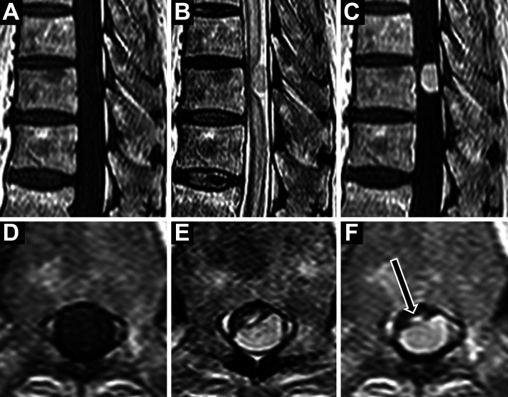
Epithelioid hemangioendothelioma (EHE) is a rare vascular tumor, and develops infrequently in the central nervous system. To our knowledge, this is the first case of EHE of the spinal cord. An 85-year-old man presented with about 6-month progressive myelopathy. Magnetic resonance imaging (MRI) demonstrated an oval-shaped intradural extramedullary mass at T10 level with extensive intramedullary edema. A reddish tumor was removed via a total laminectomy of T9-T10. Histologically, the tumor cells with nuclear atypia and active mitoses were immunopositive for vascular tumor markers, and formed a lobular architecture associated with capillary-sized vessels lined with edematous endothelial cells. Although very rare, EHE should be considered in the differential diagnosis of a spinal intradural extramedullary tumor.
Keywords: epithelioid hemangioendothelioma; spinal cord; vascular tumor.
© 2021 The Japan Neurosurgical Society.
Conflict of interest statement
Conflicts of Interest Disclosure There are no conflicts of interest and nothing to disclose.
Figures


References
-
- Marucci G, Barbanera A, Tosi AL, et al. : Epithelioid hemangioendothelioma of the spinal cord: description of a case with cytogenetic analysis. Int J Surg Pathol 14: 340– 343, 2006 - PubMed
-
- Dasgupta R, Fishman SJ: ISSVA classification. Semin Pediatr Surg 23: 158– 161, 2014 - PubMed
-
- Schattenberg T, Kam R, Klopp M, et al. : Pulmonary epithelioid hemangioendothelioma: report of three cases. Surg Today 38: 844– 849, 2008 - PubMed
Publication types
LinkOut - more resources
Full Text Sources
Research Materials