. 2022 Mar 23;96(6):e0185021.
doi: 10.1128/jvi.01850-21. Epub 2022 Jan 26.

Adenovirus-α-Defensin Complexes Induce NLRP3-Associated Maturation of Human Phagocytes via Toll-Like Receptor 4 Engagement

Affiliations

Adenovirus-α-Defensin Complexes Induce NLRP3-Associated Maturation of Human Phagocytes via Toll-Like Receptor 4 Engagement

Karsten Eichholz et al. J Virol. .

Abstract

Intramuscular delivery of human adenovirus (HAdV)-based vaccines leads to rapid recruitment of neutrophils, which then release antimicrobial peptides/proteins (AMPs). How these AMPs influence vaccine efficacy over the subsequent 24 h is poorly understood. In this study, we asked if human neutrophil protein 1 (HNP-1), an α-defensin that influences direct and indirect innate immune responses to a range of pathogens, impacts the response of human phagocytes to three HAdV species/types (HAdV-C5, -D26, -B35). We show that HNP-1 binds to the capsids and redirects HAdV-C5, -D26, and -B35 to Toll-like receptor 4 (TLR4), which leads to internalization, an NLRP3-mediated inflammasome response, and interleukin 1 beta (IL-1β) release. Surprisingly, IL-1β release was not associated with notable disruption of plasma membrane integrity. These data further our understanding of HAdV vaccine immunogenicity and may provide pathways to extend the efficacy. IMPORTANCE This study examines the interactions between danger-associated molecular patterns and human adenoviruses, and their impact on vaccines. HAdVs and HNP-1 can interact, and these interactions will modify the response of antigen-presenting cells, which will influence vaccine efficacy.

Keywords: Toll-like receptors; adenoviruses; antimicrobial peptides; innate immunity; phagocytes.


Conflict of interest statement

The authors declare no conflict of interest.

Figures

FIG 1
FIG 1
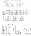
Analyses of HNP-1 binding to HAdV-C5, -D26 and -B35 by SPR. (A) Representative sensorgrams of HNP-1-HAdV interactions: HAdV-C5 (red), -D26 (black), and -B35 (blue) were coupled to the sensor chip. (B) HAdV-C5, -D26, and -B35 were covalently coupled to a CM5 sensor chip and escalating doses of HNP-1 (6.25 to 200 nM) for KD determination. Overlaid sensorgrams are shown. (C) KD of HNP-1 for HAdV-C5, -D26, and -B35. RU = resonance units.
FIG 2
FIG 2
HNP-1 interacts with HAdVs and increases infection of monocyte-derived DCs. (A) Upper panel: representative flow cytometry profiles of monocyte-derived DCs incubated with HAdV-C5, -D26, and -B35 replication-defective vectors containing fluorescent protein expression cassette. Mock-treated DCs (gray), or DCs incubated with HAdV-C5, -D26, or -B35 in the absence (red) or presence (blue) of HNP-1. Lower panel: MFI and gMFI for the FP+ and FP- cells for different capsids ± HNP-1 and fold changes in the presence of HNP-1 for a representative experiment are shown. (B) Representative flow cytometry profiles of cells incubated with HAdVs ± HNP-1. DCs were mock-treated (gray) or incubated with HAdV-C5, -D26, or -B35 alone (red); HNP-1 complexed with HAdV (blue); HAdV for 30 min and then HNP-1 (green); or HNP-1 for 30 min and then the HAdV (purple). Fluorescence was analyzed 24 h postincubation. (C) Cumulative data using HAdVs alone or HAdVs complexed with HNP-1, IVIg, or both. Two-tailed paired t-tests were used for comparison of HAdV versus HAdV + HNP-1 (n = 25, gray). Samples in black (n = 3) were used for analyses between HAdV + HNP-1 versus HAdV + HNP-1 + IVIg. (D to E) Monocytes and monocyte-derived LCs were incubated with HAdVs ± HNP-1 and transgene expression was analyzed 24 h postincubation (n = 3, statistical analyses by two-tailed paired t tests).
FIG 3
FIG 3
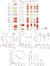
HAdV-HNP-1 induced cytokine secretion and monocyte-derived DC maturation. (A) Monocyte-derived DCs were incubated with HAdV-C5, -D26, or -B35 ± HNP-1, and cytokine secretion in supernatants was assessed by Luminex. A blanked reference is located to the left of each set of columns. For the 4 HAdV columns on the left, the reference is mock-treated cells; for the middle HNP-1, columns the reference is HNP-1-treated cells; for the “paired” columns on the right (-C5, -D26, and -B35) the reference is HAdV-infected cells compared to HAdV-HNP-1 infected cells. LPS was used as a positive control for TLR4 activation. (B) IL-1β release induced by HAdV-C5, -D26, and -B35 ± HNP-1 at 4 h (red circles) and 24 h (black dots) postincubation (n = 5). As above, cells were treated with LPS and nigericin as controls. (C) HNP-1-mediated HAdV infection of monocytes was analyzed 24 h postincubation by flow cytometry (n = 3). (D) DCs were incubated with HAdV-C5, -D26, or -B35 ± HNP-1, IVIg, or HNP-1 + IVIg. IL-1β release was quantified at 24 h postincubation (n = 3). (E) DCs were incubated with HAdV-HNP-1 complexes for 24 h. Phenotypic maturation was assessed by CD86 surface levels using flow cytometry. (F) DCs were incubated at 4°C or 37°C for 30 min with 1 mg/mL TRITC-labeled dextran, washed with PBS, fixed with 4% PFA, and analyzed by flow cytometry (lower fluorescence = lower phagocytosis = greater maturation) (n = 3). Statistical analyses were performed by two-tailed paired t tests unless otherwise noted.
FIG 4
FIG 4
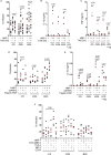
HAdV-HNP-1 complexes engage TLR4 in monocyte-derived DCs. (A) DCs were incubated with HAdV-HNP-1 complexes ± TAK-242 and analyzed by flow cytometry for vector-mediated transgene expression (n = 13). (B) IL-1β release from HAdV-HNP-1 complex-challenged monocyte-derived DCs, ± TAK-242 treatment, was quantified by ELISA (n = 3, Student's t test). (C) TNF secretion by monocyte-derived DCs following challenge with HAdV-HNP-1 complexes ± TAK-242 (n = 3). (D) Percentage of monocyte-derived DCs expressing the transgene following challenge with HAdV-HNP-1 complexes ± oxPAPC or Pepinh-TRIF (n = 6). (E) IL-1β release by monocyte-derived DCs following challenge by HAdV-HNP-1 ± oxPAPC or Pepinh-TRIF (n = 3). (F) HAdV-HNP-1 complexes were created with recombinant TLR4, TLR4/MD-2, MD-2 proteins, or anti-CD14 antibody. Recombinant protein/HAdV ± HNP-1 was then added to the DCs. Vector-mediated transgene expression was analyzed 24 h postincubation (n = 5). Statistical analyses were performed by two-tailed paired t tests unless otherwise noted.
FIG 5
FIG 5
HNP-1 induces transcription of inflammasome components. (A to C) mRNA levels of selected inflammasome components (encoded by NLRP3, CASP1, and IL1B) were analyzed using qRT-PCR 4 h postincubation. Total RNA was isolated from monocyte-derived DCs incubated with the indicated HAdV ± HNP-1; cDNAs were generated and quantified in triplicate. GAPDH mRNA levels were used to standardize samples. (D) Prior to challenge with HAdV-HNP-1 complexes, monocyte-derived DCs were treated with KCl, NAC, or MDL (n = 6). IL-1β release was quantified at 24 h postincubation. (E) Percentage of monocyte-derived DCs expressing vector-encoded transgene following KCl, NAC, or MDL (n = 3) pretreatment and subsequent challenge with HAdVs ± HNP-1. Statistical analyses were by two-tailed paired t tests unless otherwise noted.
FIG 6
FIG 6
TLR4-induced signaling and NLRP3 induction in monocyte-derived DCs. (A) DCs were treated with R406 or Bay11-7082 prior to challenge with HAdVs ± HNP-1. IL-1β release was quantified 24 h postchallenge by ELISA. LPS/nigericin was used as a control for DC activation. (B) Monocyte-derived DCs were treated with R406 or Bay11-7082 prior to challenge with HAdVs ± HNP-1. The percentage of DCs expressing the reporter gene was quantified by flow cytometry. Cumulative data from 5 donors are shown. (C) Monocyte-derived DCs were treated with MCC-950 prior to challenge with HAdVs ± HNP-1. IL-1β release was quantified 24 h postchallenge by ELISA. Statistical analyses were by two-tailed paired t tests unless otherwise noted.
FIG 7
FIG 7
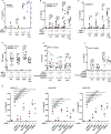
NLRP3 inflammasome is downstream of TLR4 engagement. To identify the pathway of IL-1β production, we inhibited selected caspases using (A) WEHD, (B) YVAD and VX765, and (C) Z-IEDT. IL-1β release was quantified 24 h postchallenge by ELISA. (D) DCs were preincubated with inhibitors of caspases 1/4/5 (YVAD and VX765) and then challenged with HAdV ± HNP-1. Extracellular TNF levels were quantified 24 postchallenge. (E to F) Transgene expression and IL-1β release were quantified in response to monocyte-derived DCs pretreated with RIPK1-RIPK3 inhibitors (GSK963, necrosulfonamide, or GSK872) and then incubated with HAdV-HNP-1 complexes (n ≥ 3). (G) Combined analyses of IL-1β release broken down by each HAdV type. Statistical analyses were by two-tailed paired t-tests unless otherwise noted.
FIG 8
FIG 8
IL-1β release without loss of monocyte-derived DC membrane integrity. (A) DCs were incubated with HAdV-C5, -D26, or -B35 ± HNP-1, or with LPS/nigericin. Extracellular LDH activity was quantified 4 h postincubation (n = 6). (B) 7-AAD uptake by monocyte-derived DCs, after challenge with HAdV-HNP-1 complexes, 24 h postincubation (n = 9). (C) Monocyte-derived DCs were incubated with HAdV-C5, -D26, or -B35 ± HNP-1, IVIg, or HNP-1 + IVIg. 7-AAD uptake was quantified 24 h postincubation (n = 3). Statistical analyses were by two-tailed paired t tests unless otherwise noted.

References

    1. Kremer EJ. 2020. Pros and cons of adenovirus-based SARS-CoV-2 vaccines. Mol Ther 28:2303–2304. 10.1016/j.ymthe.2020.10.002. - DOI - PMC - PubMed
    1. Lion T. 2014. Adenovirus infections in immunocompetent and immunocompromised patients. Clin Microbiol Rev 27:441–462. 10.1128/CMR.00116-13. - DOI - PMC - PubMed
    1. Kajon AE, Lamson DM, St George K. 2019. Emergence and re-emergence of respiratory adenoviruses in the United States. Curr Opin Virol 34:63–69. 10.1016/j.coviro.2018.12.004. - DOI - PubMed
    1. Lai Y, Gallo RL. 2009. AMPed up immunity: how antimicrobial peptides have multiple roles in immune defense. Trends Immunol 30:131–141. 10.1016/j.it.2008.12.003. - DOI - PMC - PubMed
    1. Nguyen LT, Haney EF, Vogel HJ. 2011. The expanding scope of antimicrobial peptide structures and their modes of action. Trends Biotechnol 29:464–472. 10.1016/j.tibtech.2011.05.001. - DOI - PubMed

Publication types

MeSH terms